section name header

Symptoms

Recurrent attacks of acute ocular pain, photophobia, foreign body sensation, and tearing. The pain may awaken patients from sleep or occur spontaneously with eye opening. There is often a history of prior corneal abrasion in the affected eye.

Signs

Critical

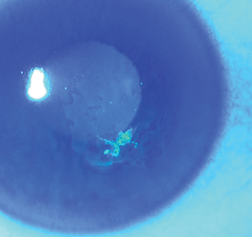
Localized irregularity with loose corneal epithelium (fluorescein dye may outline the area with negative or positive staining) or a corneal abrasion (see Figure 4.2.1). Epithelial changes may resolve within hours of the onset of symptoms so abnormalities may be subtle or absent when the patient is examined.

Figure 4.2.1: Resolving epithelial defect with paracentral area of negative fluorescein staining in recurrent corneal erosion.

Rapuano9781975243722-ch004_f002.jpg

Other

Corneal epithelial dots or microcysts, a fingerprint pattern, or map-like lines may be seen in both eyes if epithelial basement membrane (map–dot–fingerprint) dystrophy is present. These findings may also be seen unilaterally and focally in any eye that has recurrent erosions.

Etiology

Damage to the corneal epithelium or epithelial basement membrane from one of the following:

Workup

  1. History: History of a corneal abrasion? Ocular surgery? Family history (corneal dystrophy)?

  2. Slit-lamp examination with fluorescein staining of both eyes (visualization of abnormal basement membrane lines may be enhanced by instilling fluorescein and looking for areas of rapid tear break-up, referred to as “negative staining”).

Treatment

  1. Acute episode: Cycloplegic drop (e.g., cyclopentolate 1%) b.i.d. and ophthalmic antibiotic ointment (e.g., erythromycin, bacitracin) four to six times daily. Can use 5% sodium chloride ointment q.i.d. in addition to antibiotic ointment. If the epithelial defect is large, a bandage soft contact lens and topical antibiotic drops q.i.d. may be placed. Oral analgesics as needed. 

  2. Never prescribe topical anesthetic drops.

  3. After epithelial healing is complete, artificial tears at least q.i.d. and artificial tear ointment q.h.s. for at least 3 to 6 months, or 5% sodium chloride drops q.i.d. and 5% sodium chloride ointment q.h.s. for at least 3 to 6 months.

  4. If the corneal epithelium is loose or heaped and is not healing, consider epithelial debridement. Apply a topical anesthetic (e.g., proparacaine) and use a sterile cotton-tipped applicator or cellulose sponge (e.g., Weck-Cel surgical spear) to gently remove all loose epithelium.

  5. For erosions not responsive to the preceding treatment, consider the following:

    • Prophylactic medical treatment with 5% sodium chloride ointment q.h.s.

    • Oral doxycycline (matrix metalloproteinase inhibitor) 50 mg b.i.d. with or without a short course of topical steroid drops (e.g., fluorometholone 0.1% b.i.d. to q.i.d. for 2 to 4 weeks).

    • Topical azithromycin drops q.h.s. with or without a short course of topical steroid drops (e.g., fluorometholone 0.1% b.i.d. to q.i.d. for 2 to 4 weeks).

    • Extended-wear bandage soft contact lens for 3 to 6 months with a topical antibiotic and routine changing of the lens.

    • Anterior stromal puncture can be applied to localized erosions, such as in traumatic cases, outside the visual axis in cooperative patients. It can be performed with or without an intact epithelium. Stromal puncture may be applied manually with a needle at the slit lamp or with a yttrium aluminum garnet (YAG) laser. This treatment may cause small permanent corneal scars that are usually of no visual significance if outside of the visual axis.

    • Epithelial debridement with diamond burr polishing of Bowman membrane or excimer laser phototherapeutic keratectomy (PTK). Both are highly effective (up to 90%) for large areas of epithelial irregularity and lesions in the visual axis. Excimer laser ablation of the superficial stroma can be particularly helpful if repeated erosions have created anterior stromal haze or scarring. It is important to keep in mind that excimer laser PTK can lead to a hyperopic refractive shift after treatment if a deep ablation is performed.

Follow-Up

Every 2 to 5 days until the epithelium has healed, and then every 1 to 3 months, depending on the severity and frequency of the episodes. It is important to educate patients that persistent use of lubricating ointment (5% sodium chloride or tear ointment every night) for 3 to 6 months following the initial healing process reduces the chance of recurrence.