(See Figure A.6.1.)
This test distinguishes restrictive causes of decreased ocular motility from other motility disorders. One technique is the following:
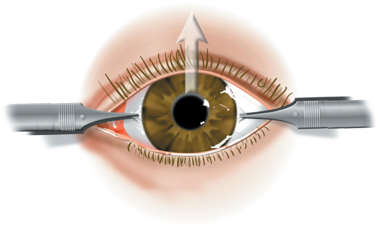
Place a drop of topical anesthetic (e.g., proparacaine) into the eye.
Use toothed forceps (e.g., Graefe fixation forceps) to firmly grasp Tenon’s close to the limbus at both locations perpendicular to the desired direction of movement. Doing so helps prevent corneal abrasions should the forceps slip. Rotate the eye in the “paretic” direction. If there is a resistance to the passive rotation of the eye, a restrictive disorder is diagnosed. This test does not require the patient to be conscious.
The patient is asked to look in the “paretic” direction while a sterile cotton swab is held just beneath the limbus on that same side. The amount of force generated by the “paretic” muscle is compared with that generated in the normal contralateral eye. The test can only be used in a cooperative, alert patient.