(See Figure 8.12.1.)
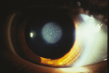
A white fundus reflex (leukocoria), absent or asymmetric red pupillary reflex, abnormal eye movements (nystagmus) in one or both eyes, and strabismus. Often appears asymptomatic in infants, delaying diagnosis. Infants with bilateral cataracts may be noted to be visually inattentive. In patients with a monocular cataract, the involved eye may be smaller. A cataract alone does not cause a relative afferent pupillary defect.
Familial: Can be autosomal dominant (most common), autosomal recessive, or rarely part of an X-linked recessive NanceHoran syndrome. Phenotype varies in terms of cataract morphology and timing of clinical onset.
Galactosemia: Cataract may be the sole manifestation when galactokinase deficiency is responsible. A deficiency of galactose-1-phosphate uridyl transferase may produce intellectual disability and symptomatic cirrhosis along with cataracts. The typical oil droplet opacity may or may not be seen. Incidence and onset of cataract may vary according to type of galactosemia (i.e., mutation of uridyl transferase, galactokinase, or epimerase). Cataract may be reversible with appropriate dietary modifications.
PFV: Usually unilateral, very rarely bilateral. The involved eye is usually slightly smaller than the normal fellow eye. Examination after pupil dilatation may reveal a plaque of fibrovascular tissue behind the lens with elongated ciliary processes extending to it. Progression of the lens opacity often leads to angle-closure glaucoma. If bilateral, 90% are associated with systemic abnormalities and require further workup.
Rubella: Pearly white nuclear cataract, salt-and-pepper chorioretinitis, microphthalmos, corneal clouding, and poorly dilating pupils. Glaucoma may occur with congenital rubella but usually does not occur in the presence of a rubella cataract. Associated hearing defects and heart abnormalities are common.
Lowe syndrome (oculocerebrorenal syndrome): Opaque lens, congenital glaucoma, renal disease, and intellectual disability. X-linked recessive. Patients mothers may have small cataracts.
Alport syndrome: Glomerulonephritis, hearing loss, eye abnormalities including cataract, anterior lenticonus, and keratoconus. Most commonly X-linked or autosomal recessive.
Others: Intrauterine infection, chromosomal disorders, aniridia, systemic syndromes, metabolic abnormalities, craniofacial syndromes, musculoskeletal disorders, and anterior segment dysgenesis.
Acquired: Trauma, drugs, uveitis, metabolic/endocrine abnormalities, radiation.
Types
Zonular (lamellar): Most common type of congenital cataract. White opacities that surround the nucleus with alternating clear and white cortical lamella resembling an onion skin.
Polar: Small opacities of the lens capsule and adjacent cortex on the anterior or posterior pole of the lens. Anterior polar cataracts usually are small and tend to grow very little over time. They may be associated with anisometropia and anisometropic amblyopia. Posterior polar cataracts are variable and may grow significantly, causing decreased vision.
Posterior lenticonus: A posterior protrusion, usually opacified, in the posterior capsule.
Posterior subcapsular: Opacification of the area immediately anterior to the posterior capsule. Most often acquired due to steroid medications, diabetes, or ionizing radiation.
History: Trauma? Maternal illness or drug ingestion during pregnancy? Systemic or ocular disease in the infant or child? Radiation exposure or trauma? Family history of congenital cataracts? Steroid use? Diarrhea is associated with cerebrotendinous xanthomatosis.
Visual assessment of each eye monocularly by using techniques for nonverbal children (Teller cards, following small toys or a light). Note briskness of pupillary response to light.
Ocular examination: Attempt to determine the visual significance of the cataract by evaluating the size and location of the cataract and whether the retina can be seen with a direct ophthalmoscope or retinoscope when looking through an undilated pupil. A blunted retinoscopic reflex suggests the cataract is visually significant. Cataracts ≥3 mm in diameter usually but not always affect vision. Cataracts <3 mm may not be inherently visually significant but have been associated with amblyopia secondary to induced anisometropia. Check intraocular pressure, corneal diameter as signs of glaucoma (see 8.13, Congenital/Infantile Glaucoma) and examine the optic nerve and retina for abnormalities.
B-scan US may be helpful when the fundus view is obscured. It is essential to rule out posterior PFV, retinoblastoma in unilateral cataract cases where the fundus is not visible.
Ultrasound biomicroscopy (UBM) can be helpful in cases of anterior segment dysgenesis or PFV.
Bilateral cataracts suggest a genetic or metabolic etiology; medical examination by a pediatrician looking for associated abnormalities is recommended.
Red blood cell (RBC) galactokinase activity (galactokinase levels) with or without RBC galactose-1-phosphate uridyl transferase activity to rule out galactosemia. The latter test is performed routinely on all infants in the United States as part of the newborn screen.
Other tests as suggested by the systemic or ocular examination. The chance that one of these conditions is present in a healthy child is remote.
Referral to a pediatrician to treat any underlying disorder.
Cataract extraction, usually within days to weeks of discovery (typically between age 6 to 10 weeks) to prevent irreversible amblyopia, is performed in the following circumstances:
After cataract extraction, treat amblyopia (see 8.5, Amblyopia).
A dilating agent (e.g., phenylephrine 2.5% t.i.d. or cyclopentolate 1% b.i.d.) may be used as a temporizing measure, allowing peripheral light rays to pass around the lens opacity and reach the retina. If the cataract is small, and the red reflex is good around the peripheral lens, this may be the only treatment needed.
Unilateral cataracts that are not large enough to obscure the visual axis may still result in amblyopia due to ansiometropia. Treat amblyopia as above.