Progressive decrease in vision, starbursts, and halos, usually beginning in adolescence and continuing into middle age. Acute corneal hydrops can cause a sudden decrease in acuity, pain, red eye, photophobia, and profuse tearing.
(See Figure 4.25.1.)
Noninflammatory ectasia of corneal stroma seen as slowly progressive irregular astigmatism resulting from paracentral (usually inferior) thinning and bulging of the cornea (maximal thinning near the apex of the protrusion), vertical tension lines in the posterior cornea (Vogt striae), an irregular corneal retinoscopic reflex (scissor reflex), and egg-shaped mires on keratometry. Inferior steepening is seen on corneal topographic evaluation, and inferocentral posterior elevation and thinning are seen on tomographic evaluation. Almost always bilateral but often asymmetric.
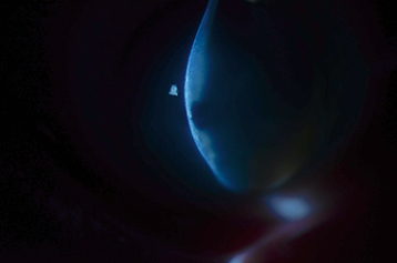
Fleischer ring (epithelial iron deposits at the base of the cone), bulging of the lower eyelid when looking downward (Munson sign), superficial apical scarring, conical reflection seen on the nasal cornea when shining a penlight from the temporal side (Rizutti sign). Corneal hydrops (sudden development of corneal edema) results from a rupture in Descemet membrane (see Figure 4.25.2).
Associations
Keratoconus is associated with sleep apnea, floppy eyelid syndrome, Down syndrome, atopic disease, Turner syndrome, Leber congenital amaurosis, mitral valve prolapse, retinitis pigmentosa, thyroid disorders, and connective tissue disorders (EhlersDanlos syndrome/Marfan syndrome). It frequently is related to chronic eye rubbing. Family history of keratoconus is also a risk factor.
Pellucid marginal degeneration: Uncommon, nonhereditary. Typically presents in the second to fifth decade. Corneal thinning in the inferior periphery from 4 to 8 oclock, 1 to 2 mm from the limbus. Absence of inflammation. The cornea protrudes superior to the band of thinning manifesting in high irregular against the rule astigmatism.
Keratoglobus: Rare, congenital, nonhereditary, nonprogressive. Uniform globular-shaped cornea with diffuse thinning from limbus to limbus. Associated with Leber congenital amaurosis, EhlersDanlos syndrome, and blue sclera.
Postrefractive surgery ectasia: After lamellar refractive surgery such as LASIK and SMILE, and rarely surface ablation, a condition very similar to keratoconus can develop. It is treated in a similar manner to keratoconus.
History: Duration and rate of decreased vision? Frequent change in eyeglass prescriptions? History of eye rubbing? Allergies? Medical problems? Family history? Previous refractive surgery?
Slit-lamp examination with close attention to location and characteristics of corneal thinning, Vogt striae, and a Fleischer ring (may be best appreciated with cobalt blue light).
Retinoscopy and refraction. Look for irregular astigmatism and an oil-drop or scissors red reflex.
Corneal topography (can show central and inferior steepening), tomography (can show posterior corneal elevation, thinning, and inferior displacement of the thinnest location), and keratometry (irregular mires and steepening).
Correct refractive errors with glasses or soft contact lenses (for mild cases) or RGP or scleral contact lenses (successful in most cases). Hybrid or piggyback contact lenses are other options.
Partial-thickness (deep anterior lamellar keratoplasty) or full-thickness corneal transplantation surgery is usually indicated when contact lenses cannot be tolerated or no longer produce satisfactory vision.
Intracorneal ring segments have been successful in getting some patients back into contact lenses, especially in mild-to-moderate keratoconus.
Corneal cross-linking (CXL) is a minimally invasive procedure performed to slow or arrest actively progressive disease by strengthening molecular bonds between collagen fibrils. The currently FDA-approved protocol involves creation of a 9-mm corneal epithelial defect after which riboflavin drops are placed on the cornea for at least 30 minutes and then ultraviolet light is applied to the cornea for another 30 minutes. While FDA approved for ages 14 years and older, it can be performed off-label for younger children. Epithelium-on protocols are under FDA investigation.
Descemet membrane usually heals and edema resolves over 3 months. However, treatment may be helpful.
Cycloplegic agent (e.g., cyclopentolate 1% t.i.d) may be beneficial for relief of pain if there is an associated anterior chamber reaction. In some patients, dilating the pupil can exacerbate photosensitivity.
Consider an aqueous suppressant such as brimonidine 0.1% b.i.d. to t.i.d.
Start sodium chloride 5% ointment b.i.d. to q.i.d. until resolved (usually several weeks to months).
Consider topical antibiotics if the epithelium is compromised to prevent secondary infection.
Consider topical steroids to suppress corneal neovascularization and/or enhance ocular comfort.
Glasses or a shield should be worn by patients at risk for trauma or by those who rub their eyes.
Intracameral air, SF6, or C3F8 may help edema resolve more quickly, but may be equivalent to conservative management in final BCVA. Such intervention can cause cataract and pupillary block glaucoma. Rarely, full-thickness corneal suturing of the cleft may speed up resolution but may also cause aqueous leakage.