Folkman, nearly four decades ago, demonstrated that tumors could not grow beyond 1–2 mm, the diffusion distance of oxygen, without the formation of new blood vessels, a process called angiogenesis.1 Angiogenesis is a critical process during embryogenesis and also during wound healing, rebuilding the endometrial lining in women after menstruation, making the placenta during pregnancy, and enabling bone and muscle growth. Angiogenesis can also be activated during pathologic conditions, such as inflammation, atherosclerosis, and agerelated macular degeneration.
Process of Angiogenesis
The body very carefully regulates angiogenesis via the “angiogenic switch.” The switch is turned on when proangiogenic factors (the most important being hypoxia) are greater than antiangiogenic factors, which oppose angiogenesis.2 When proangiogenic factors are stronger than opposing factors, the angiogenic switch is turned to the “on” position, driving angiogenesis. In the normal human, the angiogenesis switch is most often in the “off” position. For example, when cells do not get enough oxygen, they become hypoxic and release a substance called hypoxia-inducible factor (HIF), which is a transcription factor that leads to the production of vascular endothelial growth factor (VEGF). VEGF then goes to the endothelial cell, binds to the VEGF receptor (VEGFR), and starts sending a message to the cell nucleus to make more endothelial cells (proliferation) and for these new cells to migrate, forming blood vessels that will bring oxygen to the hypoxic cells. Figure 3-1 shows the process of angiogenesis. Table 3-1 presents pro- and antiangiogenic factors.
Angiogenesis is normally a very orderly, systematic process. Tissues requiring new blood vessels, such as injured tissue (e.g., wound), release angiogenic growth factors, such as VEGF, that diffuse into neighboring tissues, where they seek capillaries, and more specifically, receptors on the endothelial cells that line the nearby capillary or blood vessel. Once VEGF binds to the VEGFR on the endothelial cells, it tells the cells to proliferate and migrate. It does this by activating a signaling cascade by which this message is sent from outside the cell through the membrane of the cell (kinase domain) and then, as if by a bucket brigade, to the cell nucleus. The message tells the nucleus which genes need to be copied
Figure 3-1. Process of angiogenesis
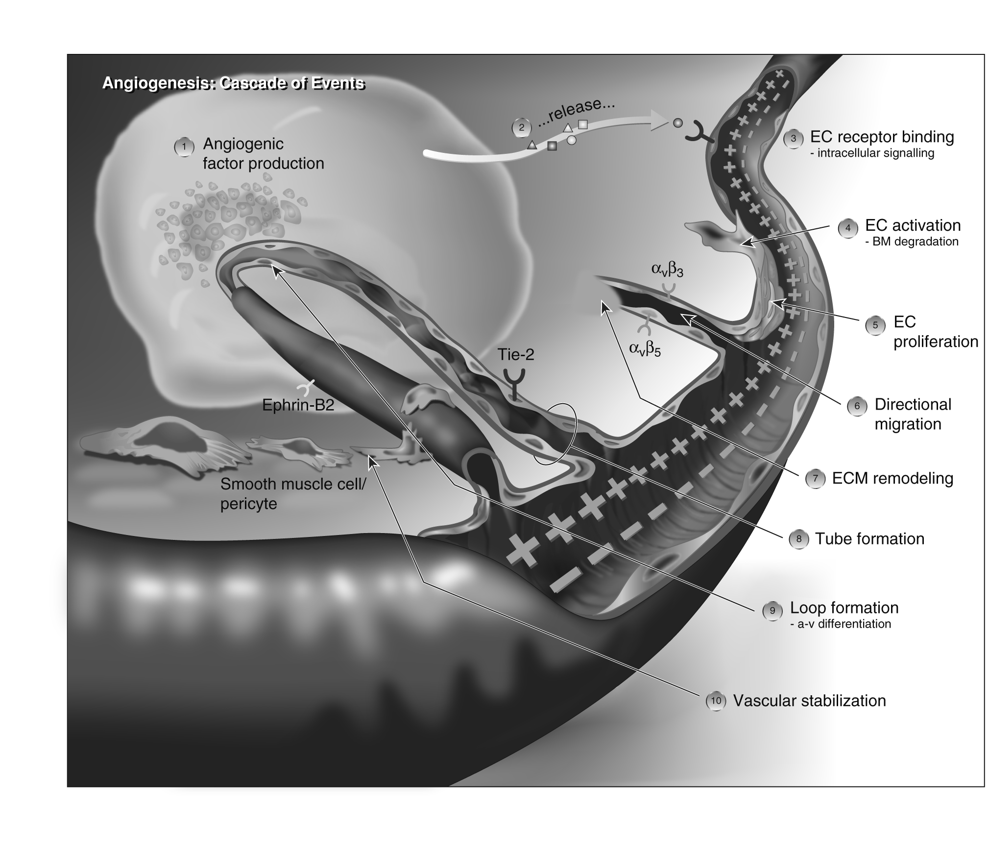
Reproduced with permission from the Angiogenesis Foundation.3
to give the protein recipes needed for cell proliferation and migration. In addition, recipes for proteins like matrix metalloproteinases (MMPs) are also copied so that the enzymes can be made. MMPs, when released, will dissolve the extracellular matrix (ECM) so that a path is made for the new and fragile blood vessel to travel to the injured tissue.
Once the recipes are copied and taken to the ribosomes to make the correct proteins, cells begin to proliferate and move through dissolved holes in the basement membrane in existing capillaries toward the injured tissue. The new cells start to sprout, and the endothelial tip cell leads the new vessel as it moves out of the existing blood vessel. The formation of the tip cell is initiated by VEGF, but differentiation is maintained by fibroblast growth factor receptor (FGFR) and VEGF signaling.4 The tip cell needs to communicate with the endothelial cells behind it, so Notch ligands delta-like 4 (DLL4) and Jagged1 become expressed on the cell, and it is via the Notch signaling pathway that the message is sent to the cells in the rear to follow the tip cell as the vessel moves toward the tumor.
| Proangiogenic factors | Antiangiogenic factors |
|---|---|
| Fibroblast growth factor (FGF), FGF-1 (acidic), and FGF-2 (basic): promote proliferation and differentiation of endothelial cells, muscle cells, and fibroblasts | Angiostatin (plasminogen fragment) |
| Vascular endothelial growth factor (VEGF): increases permeability | Fibronectin fragment |
| VEGF receptor (VEGFR) and neuropilin-1 (NRP-1, co-receptor for VEGF): promote endothelial cell survival | Heparinases Heparin hexasaccharide fragment |
| Angiopoietin 1 and its receptor Tie2: stabilize vessels | Endostatin |
| Platelet-derived growth factor (PDGF) and its receptor PDGFR: increase ECM production | Alpha, beta, and gamma interferons (IFs) |
Integrins v  3, v5, 53: bind matrix macromolecules and proteinases | Plasminogen activator inhibitor |
| Ephrin: determines whether it is an artery or a vein | Retinoids |
| Hepatocyte growth factor (HGF)/scatter factor (SF) | Metalloproteinase inhibitors (TIMPs) |
| Transforming growth factors and (TGF, TGF) | Transforming growth factor (TGF) |
| Tumor necrosis factor alpha (TNF) | Thrombospondin-1 (TSP-1) |
| Plasminogen activators: remodel ECM, release and activate growth factors | |
| Hypoxia |
Data from Kerbel RS. Molecular origins of cancer: Tumor angiogenesis. N Engl J Med. 2008;358:2039-2049; Ramanujan S, Koenig GC, Padera TP, Stoll BR, Jain RK. Local imbalance of proangiogenic and antiangiogenic factors: A potential mechanism of focal necrosis and dormancy in tumors. Cancer Res. 2000;60:1442-1448.
The tip cell has filopodia (membrane ruffles that move the cell along), and it strategizes with v3 integrin, which will pull the tip cell and its followers (stalk cells) along in the path made by membrane type 1 matrix metalloproteinase (MT1MMP or MMP14), which dissolves (remodels) the ECM in the blood vessel path, making a highway to the tumor. Under electron microscopy, the ECM looks like a jungle with many obstructions to the advancing fragile, newly forming blood vessel; thus, a discrete path needs to be created. The following stalk cells and their covering pericytes make a basement membrane and contacts between the stalk cells.
It appears that VEGF-A, which is necessary for endothial cell proliferation, migration, and survival, is also responsible for controlling the tubular sprouting of the new vessel5 (see Figure 3-1). The sprouting endothelial cells roll up to form a blood vessel tube. Integrins, specialized adhesion molecules av3 and av5, act like grappling hooks to pull the sprouting new blood vessel toward the injured tissue. The MMPs continue to dissolve the ECM in front of the advancing blood vessel so it can easily grow toward the injured tissue. Angiopoietin-1 and -2 play roles in helping the new blood vessels survive and communicate via the Tie2 receptors. They are also essential for blood vessel maturation. Blood vessel tubes connect to form blood vessel loops as shown in Figure 3-1, allowing blood from the existing blood vessel to circulate to the injured tissue.
EphrinB2 (EphB2) induces the migration of endothelial cells through the P13K pathway and helps to make adult blood vessels. EphB2 and EphB4 receptors are thought to help the endothelial cells move and form into a cord-line blood vessel; they also communicate with stromal-derived factor (SDF-1) in coordinating endothelial cell movement. SDF-1 is a chemokine that also regulates the movement of primitive stem cells. It binds to its cell surface receptor CXCR4.6 It has been implicated in breast cancer metastasis, as breast cancer cells highly express CXCR4 receptors, and common metastatic sites express their ligand CXCL12/SDF-1 alpha, such as the axillary lymph nodes and lung.7
Once formed, smooth muscle cells and pericytes give the new vessel structural support. Endothelial cells release platelet-derived growth factor (PDGF), which binds to the PDGF receptor (PDGFR) located on the cell membrane of the pericyte. Pericytes attach to the exterior of the new blood vessel, protecting the new blood vessel like the shingles of a house. Because they are located in the basement membrane of the capillaries, they serve to stabilize the walls of the blood vessel so that blood will flow smoothly. Pericytes also express receptors for PDGF- (PDGFR-) and, together with the endothelial cells that express PDGF-, help the blood vessels mature.8 The principal driver of angiogenesis in tumors is hypoxia, which causes tumors to release VEGF. The VEGF family is a subfamily of the PDGF family and has the following members: VEGF-A, VEGF-B, VEGF-C, VEGF-D (also known as FIGF), and placental growth factor. VEGF-A was discovered first and called VEGF. It is responsible
for proliferation and migration of endothelial cells, activates integrin av3, stimulates vasodilation, induces microvessel permeability, helps create blood vessel lumen, and creates the fenestrations in the blood vessel.9 VEGF-C is important in lymphangiogenesis.
Within the VEGF-A group, there are three receptors, that are called transmembrane receptor tyrosine kinases (RTKs). They include VEGFR-1 (Flt-1), VEGFR-2 (KDR/Flk -1), and VEGFR-3 (Flt-4). The different forms of VEGF all have specific receptors to which they bind. VEGF is the principal ligand for VEGFR-1 and VEGFR-2, but not VEGFR-3. VEGF-3 has VEGF-C as its ligand, and it regulates the endothelial cells in the lymphatic vessels during lymphangiogenesis.
The neuropilins (NP1 and NP2, called NRP1 and NRP2) are coreceptors for the VEGFRs, and they increase the attraction (binding affinity) of VEGF to their receptor VEGFR. Their role in cancer, however, is not clear.10 NRP1 helps integrate endothelial cell survival, together with VEGFR; it is also implicated in cell migration and invasion. NRP2 may play a role in tumorigenesis.11 VEGFR-1 is found on endothelial cells and monocytes and may modulate VEGF-2 signaling. Interestingly, VEGF has a stronger affinity to VEGFR-1 than VEGFR-2, so VEGFR-1 may act as a negative regulator of angiogenesis by preventing VEGF from binding to VEGFR-2.10 VEGFR-2 is the most well known, as it appears to mediate all the known VEGF effects.12
Within the endothelial cell, intracellular signal transduction involves similar pathways as other cancer cells. VEGF-A binds to its receptor VEGFR-1,2 on the endothelial cell. The receptors dimerize (come together), causing autophosphorylation, which activates signal transduction. Thus the receptor is turned on and a message is sent down the receptor into the cell to its internal domain tyrosine kinase. The message then travels as if by a bucket brigade (downstream) via three separate pathways to the cell nucleus13:
- The RAS→Raf→MEK→ERK pathway results in endothelial proliferation.
- The P13K →AKT pathway results in endothelial cell survival and vascular permeability.
- The p38MAPK →HSP27 pathway results in endothelial cell migration.
•Like other embryological processes that are subverted by malignant transformation, angiogenesis is also a key process in tumor formation and metastasis. Both the tumor and the stroma regulate the pathways, driving angiogenesis in cancer. VEGF is commonly overexpressed in many solid tumors.10
As illustrated by Figure 3-1, angiogenesis is an orderly series of events:
- Diseased or injured tissues produce and release angiogenic growth factors (proteins) that diffuse into the nearby tissues.
- The angiogenic growth factors bind to specific receptors located on the endothelial cells of nearby preexisting blood vessels.
- Once growth factors bind to their receptors, the endothelial cells become activated. Signals are sent from the cell's surface to the nucleus.
- The endothelial cell's machinery begins to produce new molecules, including enzymes. These enzymes dissolve tiny holes in the sheathlike covering (basement membrane) surrounding all existing blood vessels.
- The endothelial cells begin to divide (proliferate) and migrate out through the dissolved holes of the existing vessel toward the diseased tissue (tumor).
- Specialized molecules called adhesion molecules (integrins av3, av5) serve as grappling hooks to help pull the sprouting new blood vessel forward.
- Additional enzymes (MMPs) are produced to dissolve the tissue in front of the sprouting vessel tip in order to accommodate it. As the vessel extends, the tissue is remolded around the vessel.
- Sprouting endothelial cells roll up to form a blood vessel tube.
- Individual blood vessel tubes connect to form blood vessel loops that can circulate blood.
- Finally, newly formed blood vessel tubes are stabilized by specialized muscle cells (smooth muscle cells, pericytes) that provide structural support. Blood flow then begins.
Tumor cells as they proliferate need new blood vessels to bring in oxygen and glucose (food) and remove waste products.14 Hypoxia is a very strong proangiogenic factor and significantly drives new blood vessel formation. In cancer, the process of angiogenesis is very similar to that depicted in Figure 3-1 and is driven by hypoxia and local oncogenes. However, the blood vessels that are formed are very different. Key targets include VEGF, VEGFR-1 and -2, angiopoietin-1 and -2, Tie2, the integrins that pull the new blood vessel sprout toward the tumor, MMPs, and the pericytes. The hypoxic tumor cells release factors, such as VEGF-A, thus activating the endothelial cell via the VEGF receptor (VEGFR-1 and -2) and starting a process called “sprouting angiogenesis.” As we have seen in normal angiogenesis, the activated endothelial cell starts proliferating and then releases proteases (enzymes) that dissolve the basement membrane of the blood vessel so the new endothelial cells can migrate from the vessel and make a new blood vessel tube. These new endothelial cells migrate out of the holes in the basement membrane into the surrounding extracellular matrix and make solid sprouts that link to the existing blood vessel and pull the sprout toward the tumor (source of growth factors). As the endothelial cells migrate to form the blood vessel tube, they are pulled along by integrins (adhesion molecules) that pull the tube along like grappling hooks. The sprouts are linked to make loops, the loops pull together to form a complete tube, and blood begins to flow to the tumor.
However, the formed blood vessels are very different. Tumor blood vessels are poorly made. They are characterized by highly branched and tortuous vessels, with some vessels being very narrow while others are dilated. Some branches lead to dead ends; others are so narrow that blood cannot reach parts of the tumor and it necroses. This poor vessel construction may be due to the antagonistic relationship between angiopoietin-1 and angiopoietin-2. Together, these molecules stimulate the endothelial cells to recruit pericytes and smooth muscle to the newly forming blood vessel, as this is necessary for the blood vessel to mature and survive. If there is an imbalance in the angiopoietin-1 and -2, together with a high level of VEGF, poor construction of the tumor blood vessels may result. The vessels are highly permeable, with fenestrations (or windows) in them. This permeability is caused by VEGF (which when first discovered, was called “permeability factor”), and it allows fluid to leak into the interstitial spaces of the tumor and raise interstitial pressure. Often, chemotherapy does not reach the tumor due to high interstitial pressure in the tumor, as well as the blinded, tortuous, and narrow blood vessels. Hypoxic tumor cells become resistant to the administered chemotherapy. Microvessel density (MVD) is a surrogate for the extent of tumor angiogenesis.15 Tumors with high MVD are usually aggressive, and predictive of early recurrence, high potential for metastasis, and poor survival.16 Antiangiogenic drugs work by preventing new blood vessel formation and normalization of existing vasculature so that the interstitial pressures in the tumor no longer prevent the entry of chemotherapy.
In the stroma, tumor-associated macrophages (TAMs) and proinflammatory molecules help to drive angiogenesis. In fact, it appears that angiogenesis begins in the stroma long before the basement membrane has been broken down.17 Cross-talk between tumor cells, endothelial cells, and stromal cells is mediated by the Notch pathway, stimulating tumor angiogenesis.18 Notch signaling is activated when the bordering endothelial, tumor, and stromal cells touch each other (cell-to-cell contact) between the Notch receptor, which is on the endothelial cell, and its ligand. Its ligand, either Jagged1 (which is strongly proangiogenic), or delta-like 1 (DLL1) or delta-like 4 (DDL4) (which are negative regulators of angiogenesis), is present on the tumor cell. If Jagged1 binds to Notch, this activates the Notch pathway and starts the proliferation and differentiation of the endothelial cells, in concert with VEGF. For example, Jagged1 is found expressed in squamous cell head and neck cancer cells and is induced via the mitogenactivated protein kinase pathway by hepatocyte growth factor (HGF, scatter factor), EGF, and TGF, which not only drive angiogenesis but also tumor cell proliferation and progression.19 DLL4 controls the degree of sprouting by turning down the activity (downregulation) of VEGFR-2. The endothelial cells of tumor blood vessels highly express the Notch ligand DLL4, and it is controlled by VEGF. High levels of VEGF cause increased expression of DDL4 found in the endothelial tip cell or the endothelial cells growing in the hypoxic tumor center.20
The Notch signaling pathway is very complex. Research continues to try to refine our understanding of the many roles, relationships, and interactions with other signaling pathways so effective targeted agents can be developed.21 Not only can tumor and tumor endothelial cells activate Notch signaling, but so too can inflammatory cells.
In addition, tumors can make their blood vessels by having the myofibroblasts in the tumor-associated stroma send chemotactic signals (e.g., stromal cell-derived factor [SDF-1], CXCL12), which will recruit precursors of endothelial cells in the stroma.17 VEGF is also released by cancer cells, TAMs, and fibroblasts in the tumor-associated stroma, which helps the endothelial cells mature into adult, functional endothelial cells that can make blood vessels.22 Tumor cells clearly can produce VEGF via epidermal growth factor receptor (EGFR) signaling, but macrophages and myofibroblasts can as well. TAMs release powerful proangiogenic cytokines and growth factors, in addition to VEGF, such as TNF, IL-8, and basic fibroblast growth factor (bFGF).23 TAMs also express proteases such as urokinase-type plasminogen activator and the matrix metalloproteinases MMP-2, MMP-7, MMP-9, and MMP-12 that will make the path for the new blood vessel as it grows toward the tumor. Inflammatory cytokines promote tumorigenesis (malignant transformation) by stimulating tumor cell activation and cancer stem cell proliferation.24
A proangiogenic force, bFGF binds to its receptors on the endothelial cell. The endothelial cells start to proliferate. Angiopoietin-2 apparently destabilizes quiescent endothelial cells and may contribute to the new vessel sprouting. One of the endothelial cells becomes a tip cell, probably induced by VEGF, as discussed. As released enzymes dissolve holes in the basement membrane, the endothelial cells migrate out to the ECM. MMPs dissolve the ECM so the developing blood vessel tube can push its way through the tissue layers toward an area of the highest concentration of angiogenic factors (the tumor), which include bFGF, TGF, IL-8, angiopoietin, angiogenin, and PDGF.17 Specific integrins help regulate angiogenesis and lymphangiogenesis. They help the endothelial cells migrate and survive during the process of new growth. They are also excellent targets for anticancer therapy.
Some TAMs have Tie2 expression, which gives them a proangiogenic ability. Tie2 is an RTK found on the developing vascular endothelial cells.22 27 Tie receptors and their angiopoietin-1 and -2 ligands are important in vascular remodeling.25 In particular, angiopoietin-1 and its receptor Tie2 play a significant role in regulation of capillary-like tubule formation and survival of endothelial cells.26 Angiopoietins appear to be very important in angiogenesis. They modulate Tie2–TAM activity and help build efficient pericytes on the vessel walls. As the blood vessel normally develops, pericytes are attracted to the new vessel by PDGF/PDGFR; they are then tacked on in tight formation to the outside surface of the vessel like shingles to protect the vessel. The pericytes secrete angiopoietin-1, which binds to Tie2 (RTK) on endothelial cells, making the pericytes stick tightly to the vessel exterior. In cancer, the pericytes are fewer and are
very loosely attached to the blood vessel exterior; in some cases, they may even be absent. This contributes to the leakiness of tumor blood vessels. Because cancer metastasizes by entering the blood or lymph vessels, these blood vessels with fenestrations (made by VEGF) and loose vessel exterior (few pericytes) help the tumor to metastasize. Interestingly, in inflammation, angiopoietin-1 stabilizes vessels, decreases leaking from the blood vessels, and decreases the number and size of gaps between adjacent endothelial cells.
Angiopoietin-2 was originally thought to be a Tie2 receptor antagonist, as it binds to Tie2 but does not always activate Tie2 receptor signaling. However, it does compete with angiopoietin-1, blocking angiopoietin-1 from binding to Tie2 and, in effect, turning it off. Angiopoietin-1 is expressed by pericytes and smooth muscle cells. In contrast, angiopoietin-2 is expressed by activated endothelial cells where active remodeling is taking place as it destabilizes endothelial cells.28 In addition, it appears that angiopoietin-1 and -2 are both necessary for maturation of blood and lymph vessels.29 Thus, there appears to be a critical balance between angiopoietin-1 and -2 in tumor angiogenesis.30
Once the tumor becomes invasive, breaching the basement membrane and entering the stroma, angiogenesis begins in earnest. In addition, as one wonders what the immune system is doing to halt tumor growth, it is important to note that VEGF has another role to play. Normally, when the body recognizes an antigen that should not be there, it calls in immature dendritic cells from the bone marrow and peripheral tissues to the antigen. The dendritic cell then engulfs the antigen, which activates the dendritic cell and causes it to differentiate into a mature dendritic cell. The mature dendritic cell presents the antigen to the immune system (in the lymph nodes, CD4 and CD8 T cells) so that any microorganisms with this antigen can be rounded up and killed. It is important in cancer that the dendritic cells not be allowed to mature, as they would otherwise find the antigens from cancer cells in the microenvironment that have died. VEGF and other factors made by the tumor recruit the immature dendritic cells and then prevent them from maturing; the immature dendritic cells then try to present the tumor antigens to the immune system and are ineffective. The failure of the dendritic cells gives the tumor with this antigen “immune privilege” so it will not be attacked by the immune system.10
In addition, tumors secrete VEGF that attracts via chemotaxis, bone marrowderived cells, including hematopoietic and endothelial cell precursors that have VEGFR-1 receptors. In the metastatic niche, VEGFR-1–positive cells may migrate to the organ that will host the new metastatic cells and then attract bone marrowderived cells that express VEGFR-1 to the new metastatic niche. They can begin making the stroma so that when the cells arrive, blood vessels will be ready to nourish the new tumor. In addition, endothelial progenitor cells expressing VEGFR-2 are then incorporated into the growing vascular bed to construct the blood vessels in the stroma and tumor.10