A second important pathway that affects angiogenesis is the mTOR, so named because rapamycin was found to inhibit the molecule, and the mTOR molecule was a target of rapamycin. Unfortunately, rapamycin is very immunosuppressive and, therefore, has not been successful in anticancer strategies. Clearly, blocking mTOR complex 1 would turn off cell proliferation and survival, and the search has been underway for analogues (or “rapalogues”) of rapamycin (sirolimus). Of note, mTOR is highly conserved, and all organisms possess it. In the embryo, if mTOR is lost, the embryo dies.48 Figure 3-3 shows the mTOR signaling pathway.
mTOR plays a central role in angiogenesis, as it controls production of HIF proteins HIF1a and HIF1b. HIF is a critical transcription factor that allows the expression of genes whose products (proteins) are involved in angiogenesis, as well as cell proliferation, motility, adhesion, and survival—all qualities necessary for tumor progression.50 HIF induces gene expression to produce VEGF (which stimulates endothelial cells to proliferate and migrate toward the tumor in a new tube) and angiopoietin-2 (which destabilizes existing blood vessels so they can develop the tube extension in concert with VEGF and then grow toward the tumor). Angiogenesis is tightly controlled. Hypoxia tips the balance toward angiogenesis to increase the oxygen delivered to tissues and is mediated by HIF, which is controlled by the von Hippel–Lindau (VHL) protein, a tumor suppressor. During hypoxia, HIF is released so new blood vessels can be stimulated, and when tissue is oxygenated, HIF is rapidly broken down. In the presence of very high levels of HIF, apoptosis occurs.50 When tumors subvert the upstream signaling pathways, mTOR is activated, and high levels of HIF are produced; if VHL protein is mutated or lost, HIF is not broken down, and high levels persist. This
Figure 3-3. mTOR signaling in epithelial cells
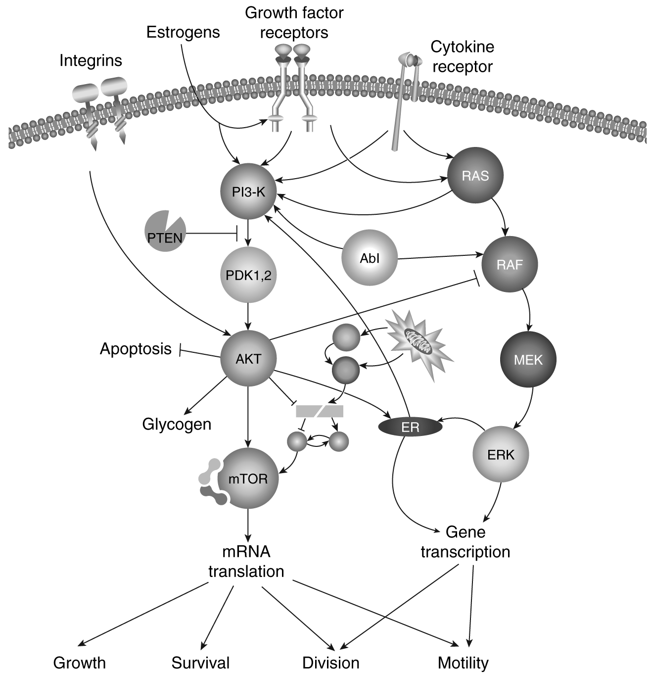
Modified from Novartis Oncology.49
way the tumor can get the proteins and essential nutrients it needs to continue to proliferate and have adequate energy even in hypoxic conditions, such as aerobic glycolysis.51 Activated mTOR also upregulates VEGF-C, which is associated with lymphangiogenesis.52
This important cell-regulating pathway can be thought of as a Grand Central Station of many critical cell functions, such as sensing the environment and helping the cell respond to these changes (e.g., insufficient nutrients, stress), but it also regulates the key proteins that are involved in cell growth, movement, death (apoptosis), and division, as well as replication of DNA. One of the proteins that it controls is VEGF-A. mTOR controls the cells' response to hypoxia by transcribing VEGF-A in response to HIF-1 , as well as a number of other hypoxia-related proteins that tumors express.49 We have already discussed the
important role of VEGF-A (or just VEGF) in initiating and continuing the growth of new blood vessels to supply tumor growth beyond 1–2 mm. mTOR is considered a biologic switch. It senses the availability of nutrients (e.g., amino acids, glucose, and energy) that the cell will need to continue to survive. It processes growth signals from other cells, such as hormones or growth factors. It also processes and assesses the threat of stressors that may cause the cell to die, such as hypoxia, DNA damage, heat shock, and oxidative stress. It is like a large computer or trafficking center that decides what the cell should do based on the given variables and switches on or off the process of cell growth, motility, division, and death accordingly. If the cell does not have enough nutrients or energy to divide and the cell receives messages to divide, the cell will either stop cell division (e.g., restriction point R1 in the G1 phase of the cell cycle), or it will turn on a process called autophagy, which allows the cell to break down some of its own internal cell organelles to free up the elements to make the required components the cell needs. If the cell senses hypoxia, the gene for VEGF-A is expressed, the gene is transcribed by mRNA, which carries the recipe for the protein to the ribosomes in the cytoplasm of the cell, and VEGF-A is made and released to start the mTOR signaling in the process of angiogenesis.
One may wonder, how does mTOR, a serine/threonine protein kinase found in the cytoplasm of the cell, get the messages to help it make the decisions about regulating cell growth, proliferation, motility, and survival, as well as making the proteins it needs to regulate these pathways? It gets the messages from receptors on the outside of the cell (upstream) such as insulin-like growth factors (IGF-1, IGF-2), EGFR, HER2, VEGF, and PDGF, as well as cytokine receptors on the cell surface. Nutrients also weigh in here—the presence or absence of amino acids and glucose, as well as the level of oxygen. Also important is the cell's adhesion status, which is given to it by integrins on the cell surface. In cancer, many of these receptors are overexpressed, turning on the mTOR machinery so that the cell proliferation and angiogenesis occur without interruption. Messages from inside the cell, such as from estrogen, can also turn on the mTOR machinery. Much like the signal transduction cascades already discussed, the message from outside the cell reaches the mTOR molecule by a series of bucket brigade-like steps, each involving phosphorylation of enzymes by protein kinases along the cascade. Again in cancer, as the proto-oncogenes are mutated to form oncogenes that drive cell division, and as tumor suppressor genes are silenced, mTOR can receive continuous signals to switch the cell machinery “on” (see Figure 3-3).
Many of the genes mutated in cancer are upstream of mTOR and are responsible for control of the mTOR pathway. The silencing of the tumor suppressor gene PTEN (phosphatase and tensin homologue) activates mTOR signaling. PTEN is often deleted from chromosome 10.
Mutations in the RAS signaling pathway: Normally, the ras gene is a proto-oncogene, and it codes for proteins that bring the message from the cell surface growth factor receptors to other protein messengers further down the signal cascade as part of the bucket brigade. It lies inside the cell surface, in the cytoplasm. Once RAS gets the message to help the cell grow or divide, the next step is raf; through phosphorylation, raf is activated, and it then sends the message downstream to MEK. MEK is phosphorylated and sends the message downstream to MAP kinases, which are also called the extracellular signal-regulated kinase or ERK pathway. As shown in Figure 3-4, ERK sends the message into the nucleus, resulting in gene transcription; thus, the proteins are made that make the cell undergo cell division and develop motility. However, there is much cross-talk with the RAS/ERK pathway, such as RAS with P13K (phosphoinositide 3-kinases), which then leads to stimulation of mTOR. RAF talks with Abl kinase, which talks to P13-K. Unfortunately, in many tumors, the RAS protooncogene is mutated, forming the ras oncogene, such as in cancers of the pancreas, colon, thyroid, and lung. This mutation results in increased signaling along this pathway (gain of function); the RAS oncogene is turned on and keeps sending messages to the cell nucleus instead of stopping after the message is delivered as happens in the normal cell.
Mutations in the P13K/AKT signaling pathway: Normally, P13K transduces (sends) the message it receives from activated receptors on the outside surface of the cell, or from RAS, downstream to the cell nucleus via mTOR. For example, it is involved in getting the message from the insulin-like growth factor (IGFR) to regulate glucose uptake in the cell. P13K signaling leads to cell growth, survival, proliferation, and motility through mTOR. P13K is mutated in many cancers, such as some colon, gastric, breast, ovarian, and lung cancers and glioblastoma. In addition, P13K also talks with PTEN, a tumor suppressor gene, which antagonizes P13K and thus can reverse its activity. PTEN is silenced (mutated) in many tumors as well. These tumors that have mutated PTEN include glioblastoma, prostate, and endometrial cancers. These mutations are thought to be linked to tumor insensitivity to insulin and the availability of nutrients (calories).53 In the lab, if either a normal P13K or PTEN correction is made, then the tumor cell becomes dietary-restriction sensitive. PTEN mutation also allows cells to survive when they detach from the basement membrane (anchorage independence); in normal cells, detachment and migration to another area, except for certain leucocytes, is met with apoptosis. In patients with breast cancer who are HER2+, those with mutated PTEN (thus silencing PTEN) become resistant to trastuzumab.54
When P13K is activated, it is phosphorylated and sends the message to the next step in the bucket brigade. That molecule then phosphorylates and sends the message to AKT. AKT is also a serine/threonine kinase and is also known as protein kinase B (PKB). Once it receives the message to grow, survive, proliferate,
Figure 3-4. Multiple signaling pathways
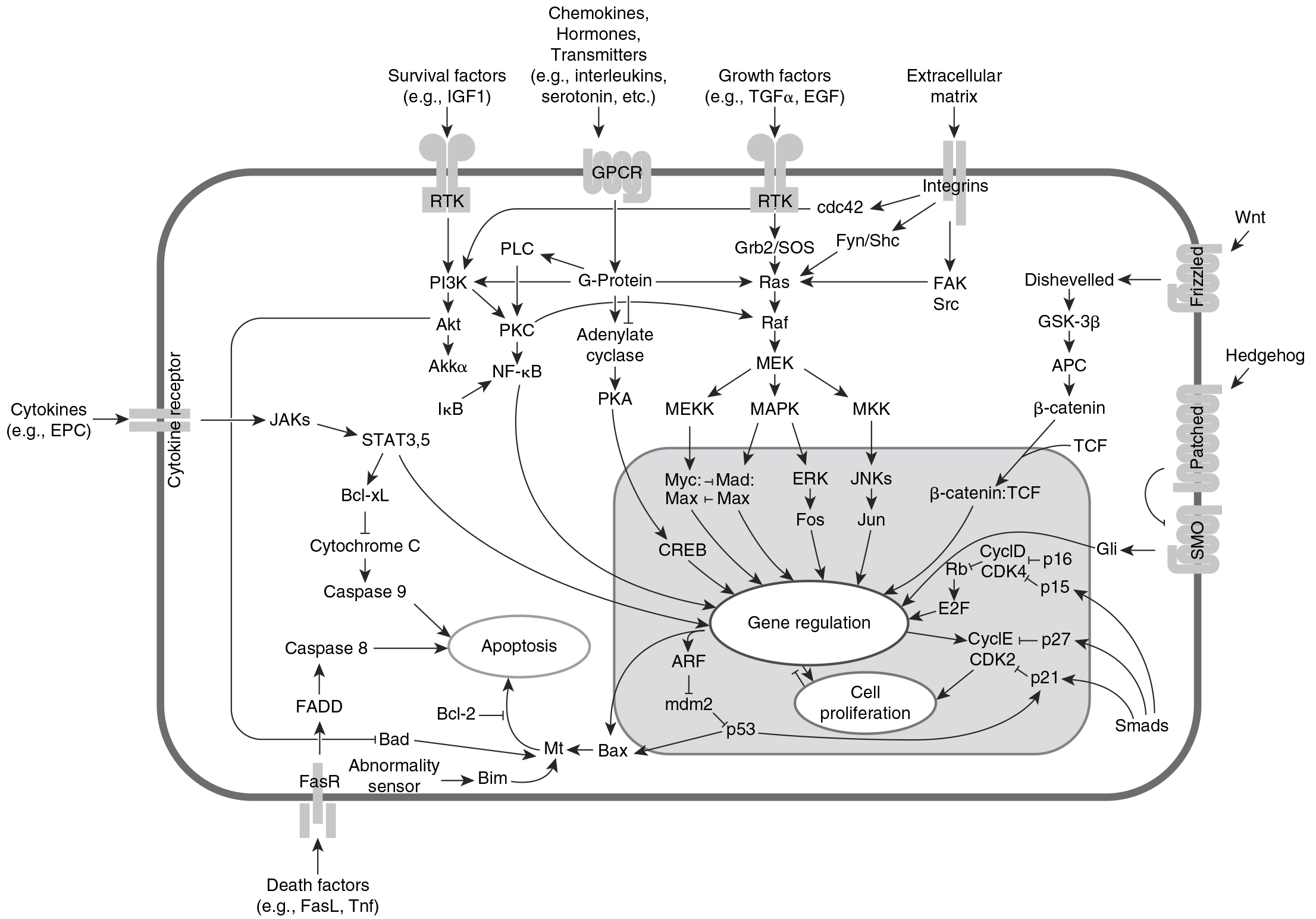
or move, it sends it along to the cell nucleus for action (e.g., gene transcription). There are three family members, and two are well known. Akt1 promotes survival of the cell by blocking apoptosis. It is very important in balancing the proand antiapoptotic forces in favor of cell survival.55 It can induce protein synthesis as well. Akt2 is important in the insulin signaling pathway, as it pays attention to the glucose level in the cell and is able to induce glucose transport to make glycogen stores.
AKT is frequently mutated or activated (i.e., becomes an oncogene) in cancer, and high levels have been found in tumors of the gastrointestinal (GI) tract, prostate, and breast.56 High levels of Bcl-2 may predict resistance to an mTOR inhibitor.57
mTOR and angiogenesis: Cancers that overexpress HIF- are often aggressive, associated with a poor prognosis, and have mutations in the mTOR signaling pathways. When hypoxia activates mTOR signaling, hypoxic stress genes are turned on, and proteins like VEGF and PDGF are synthesized to relieve the hypoxia by providing new blood vessels. These ligands bind to their respective receptors on the endothelial cell and pericyte, begin proliferation and migration, and orchestrate the building of new tumor blood vessels.
mTOR inhibitors block angiogenesis by preventing the release of VEGF and PDGF, thus directly decreasing angiogenesis. In addition, mTOR inhibition blocks the growth and proliferation of both tumor and endothelial cells as the synthesis of necessary proteins is halted. This blocks tumor cell proliferation and makes tumor cells undergo apoptosis.
Immunomodulatory agents: As discussed, the microenvironment is very important in angiogenesis. It is unclear how the immunomodulatory drugs thalidomide and lenalidomide exert their antiangiogenic properties. These drugs inhibit proinflammatory cytokines, TNF, IL-1 , and IL-6. One study by Dredge et al. showed that lenalidomide inhibited endothelial cell migration and Akt phosphorylation, leading to antiangiogenesis. However, it did not affect endothelial cell proliferation.58