The appearance of a cell or phenotype is determined by which genes are expressed (“turned on” or transcribed), such as blue eyes in a blue-eyed person. Outside the DNA, genes in the DNA can be turned on or off by remodeling the chromatin (which is DNA plus proteins). The chromatin can be remodeled by two epigenetic processes. One way is by modifying the histones around which the DNA is coiled, and the second is by adding or removing methyl groups from specific genes in the DNA. Histones are structural proteins that pack the DNA into a tightly wound double helix or chromatin complex so that the DNA coils around the histones. Histones and methylation also are able to change the DNA so that once a cell has become specialized or differentiated, it cannot go back to being a stem cell. Histones do this by becoming modified, then becoming incorporated into the DNA strand, and finally altering the shape of the histones around the gene(s) that have brought about the differentiated state. It is now thought that histone modification may be influenced or mediated by small, noncoding RNAs (see Chapter 2).
First, because the DNA is wrapped around the histone proteins, the proteins are in close contact with the genes on the DNA strands. Normally, a cell controls the coiling and uncoiling of the DNA strand around the histones when it wants to turn a gene on (uncoil it) or turn it off (coil it). It does this by using histone acetylase to acetylate a section of the histone (lysine residues) to release the tension and loosen the chromatin or DNA so that it can open the DNA strands to be copied. If the chromatin structure is closed (i.e., if the DNA is so tightly coiled that the DNA cannot open to allow m-RNA to come close and copy or transcribe a gene), then that gene is essentially shut off or silenced. The cell closes the chromatin structure by using HDAC, which removes an acetyl group from the lysine residues and makes the DNA tightly coil or condense. There are four classes of HDACs. Some malignancies cause excessive HDACs, closing the chromatin or effectively silencing certain genes. When this process affects a TSG, the brakes are removed from cell division. If it affects a cell cycle protein that normally turns off the cell cycle, then again, the gene is silenced and the cell cycle is left in the “on” position, allowing any cell through to divide.
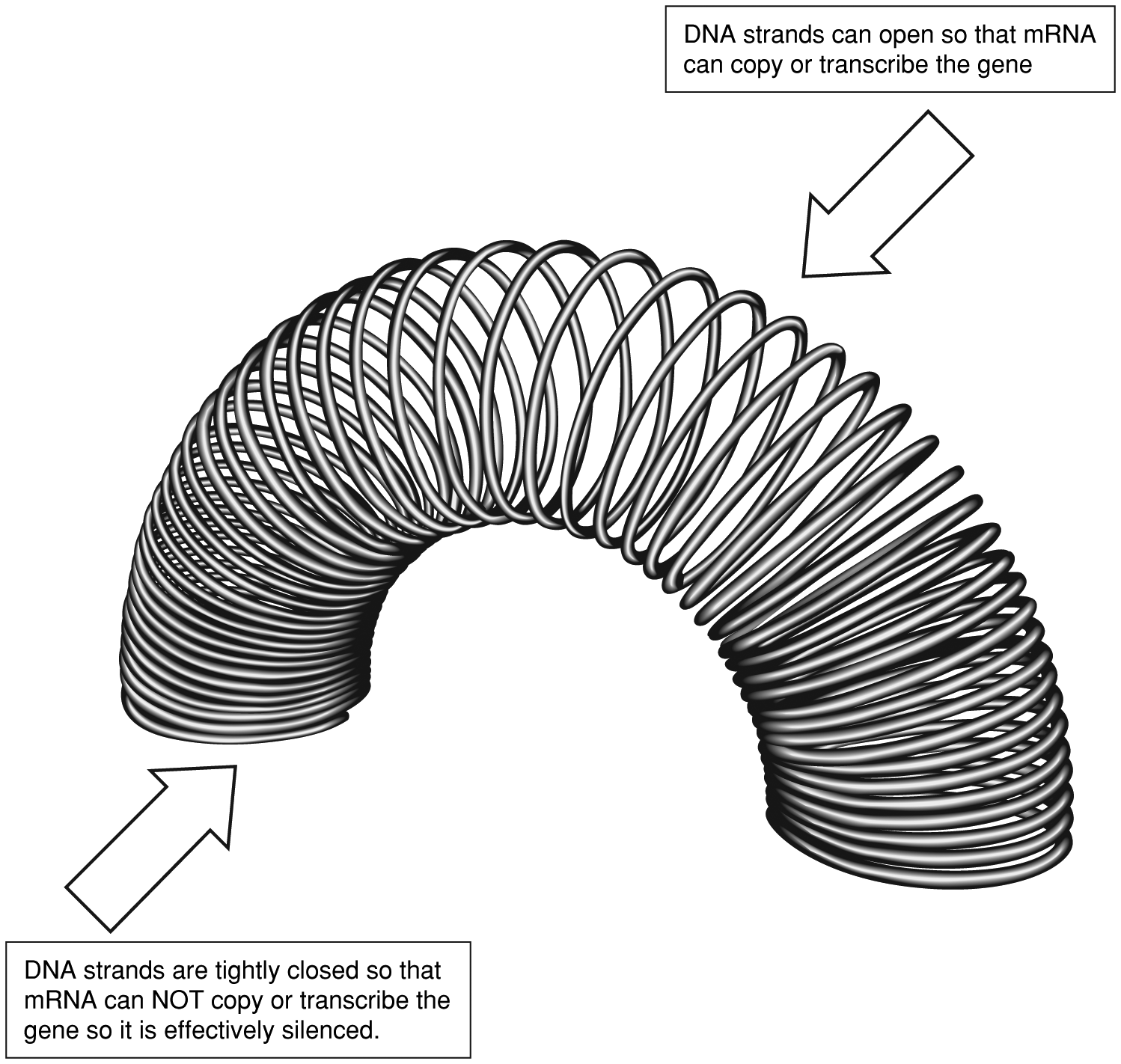
Another way to think of this is as a slinky toy (Figure 1-6). When the slinky is against a flat surface, the rings are closed. When the slinky moves, the rings at the top open. Similarly, the DNA strands need to pull apart like the open slinky
Figure 1-6. Histones are wrapped around DNA, and they influence gene expression like a slinky. When the slinky is open, it expresses the gene; when it is closed, the gene is turned off
Data from Wilkes GM. Drug essentials: Histone deacetylase inhibitors. ONCOLOGY Nurse Edition 2007;21(11):39–40; Garber K. Silence of the genes: Epigenetics arrives. J Natl Cancer Inst. 2002;94(11):793–795.
for mRNA to slip in and copy the gene sequence and to take this copy to the ribosome in the cytoplasm. If the DNA strands are tightly coiled and closed, like the end of the slinky on a flat surface, then mRNA cannot get in to transcribe the gene, and essentially the gene is shut off. The net effect is that the gene is not effective in producing proteins that will stop malignant cell division, even though the TSG is not mutated.
Cancer treatment has progressed in finding agents that inhibit HDACs. Inhibiting HDACs causes increased histone acetylation, resulting in uncoiling of the DNA from the tightly wound chromatin and in the ability of the TSGs to be transcribed or “turned on.” This slight alteration in the transcription of a few genes results in cell cycle arrest, differentiation and/or apoptosis, and other lesserknown effects that cause cancer cell death.57 59 Initially, investigators found that HDAC inhibitors can turn on the gene expression of p21, which regulates p53 activity, a very important TSG that is silenced in many solid tumors.60 More recently, vorinostat (Zolinza), an HDAC inhibitor that inhibits class I (HDAC1, 2, 3) and class II (HDAC6) HDACs, was approved by the Food and Drug Administration (FDA) for treatment of patients with refractory cutaneous T-cell lymphoma.
The second way the cell can modify gene expression epigenetically is by modifying the amount of methylation in the DNA of the cell. This modification can be
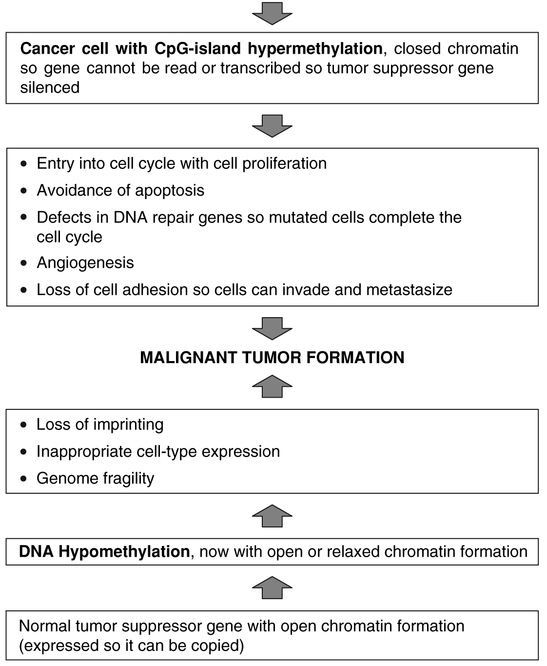
done in two ways, and each way involves different DNA sequences61 (Figure 1-7). In the first of these, methyl groups are added to the DNA in areas that are normally unmethylated. These methyl groups are added at sites characteristically called CpG sites, which change cytosine to 5-methycytosine. The 5-methycytosine is then incorporated into the DNA strand because it behaves like cytosine alone. Areas of DNA that have many methyl groups or that are hypermethylated are silent, and the genes in this area are not transcribed.
Unfortunately, in cancer cells, these areas code for TSGs. DNA methylation patterns are influenced by the environment, and this can be inherited. In cancer, TSGs are often silenced, and CpG islands are often found with promoters of cancer-associated genes.62 63 Hypermethylation leads to entry into the cell cycle, avoidance of apoptosis, defects in DNA repair, angiogenesis, and loss of
Figure 1-7. Comparison of methylation
Data from Esteller.62
cell adhesion.62 TSGs known to be hypermethylated and thus silenced in cancer include:
- MLH1—DNA mismatch repair, in colon, endometrium, and stomach, causing frameshift mutations
- BRCA1—DNA repair, transcription, in breast and ovary, causing double-strand breaks
- p16INK4a—Cyclin-dependent kinase inhibitor, in many cancers, allowing entrance into cell cycle
- p15INK4b—Cyclin-dependent kinase inhibitor in leukemia, allowing entrance into cell cycle
- p14ARF MDM2 (mouse double minute 2)—Inhibitor, in colon, stomach, and kidney cancers, causing degradation of p53 (the master policeman of the genome)
Cancer-associated hypermethylation of promoter regions of genes that protect against cancer is more common than cancer-associated hypomethylation that activates cancer genes involved in tumor formation, invasion, and metastasis. This second process of hypomethylation is less understood. In general, the DNA genome in cancer cells is hypomethylated compared to normal cells. In a study by Estécio et al. of colon cancer cells, those that showed MSI were hypermethylated compared to similar cells without MSI, which remained hypomethylated.64 The areas in cancer cells that are hypomethylated are DNA sequences that are highly and moderately frequently repeated, which suggests an independent role in tumor formation and progression.61 Cancer-associated hypomethylation appears to be related to loss of imprinting (certain genes are expressed based on the parent, such as expression of the gene in the copy or allele from the mother, such as CDKN1C, or from the father, such as IGF-2), inappropriate cell-type expression, genome fragility, and activation of sequences that lead to tumor formation. CDKN1C is a cyclin-dependent kinase inhibitor implicated in cancer,65 and IGF-2 is implicated in the formation of neuroblastomas.66 It also raises a flag of caution when hypomethylating drugs such as 5-azacytidine (Vidaza) are used therapeutically. Vidaza is FDA indicated for the treatment of patients with specific myelodysplastic subtypes or chronic myelomonocytic leukemia.
In addition, DNA methylation can also affect the length of the telomere, or the end of the chromosome, which is normally shortened with each cell division. After about 60–70 cell divisions, the chromosome is too short, and the cell is sent to programmed cell death, called senescence. In the embryo, and in up to 90% of malignant tumors, telomerase, the enzyme that lets the cell replace the snipped-off portion of the chromosome so that the length is not altered, is found in high amounts.30 67 Telomeres are formed by repeated areas of guanine-rich sequences (TTAGGG DNA repeats). However, for the remaining tumors without telomerase, Blasco found that telomerase activity can be regulated by promoter
methylation, and Vera et al.68 showed that although telomeric DNA repeats cannot be methylated, the subtelomeric DNA repeats can.69