The microenvironment is critical to cancer's successful transformation, invasion, and metastasis, just as the seed needs fertile soil. Angiogenesis appears to be a key process during all phases of malignant growth. This process and the corresponding agents are discussed in Chapter 3. In particular, the immunomodulatory thalidomide derivative agents exert their effect on the microenvironment. While much has been learned about the multiple signaling pathway in cancer formation and invasion, there remain many questions about metastasis. One exciting advance has been in the understanding of bony metastasis from patients with advanced breast and prostate cancers and multiple myeloma. Unfortunately, bony metastasis can compromise quality of life through limitation of mobility (e.g., related to pathological bone fractures), pain, and life-threatening hypercalcemia.1 Also, it is a frequent occurrence and may affect up to 70% of patients with breast or prostate cancer.2 The process for each of these malignancies will be reviewed. Although the discussion will be simplified, it is important to remember that the process is very complex; for example, women with estrogen-positive breast cancer have frequent metastases to bone, while women with estrogen-negative breast cancer more commonly have visceral metastases, such as to the liver.3 In addition, tumors in the lung or colon or renal cell cancer also metastasize to the bone.4
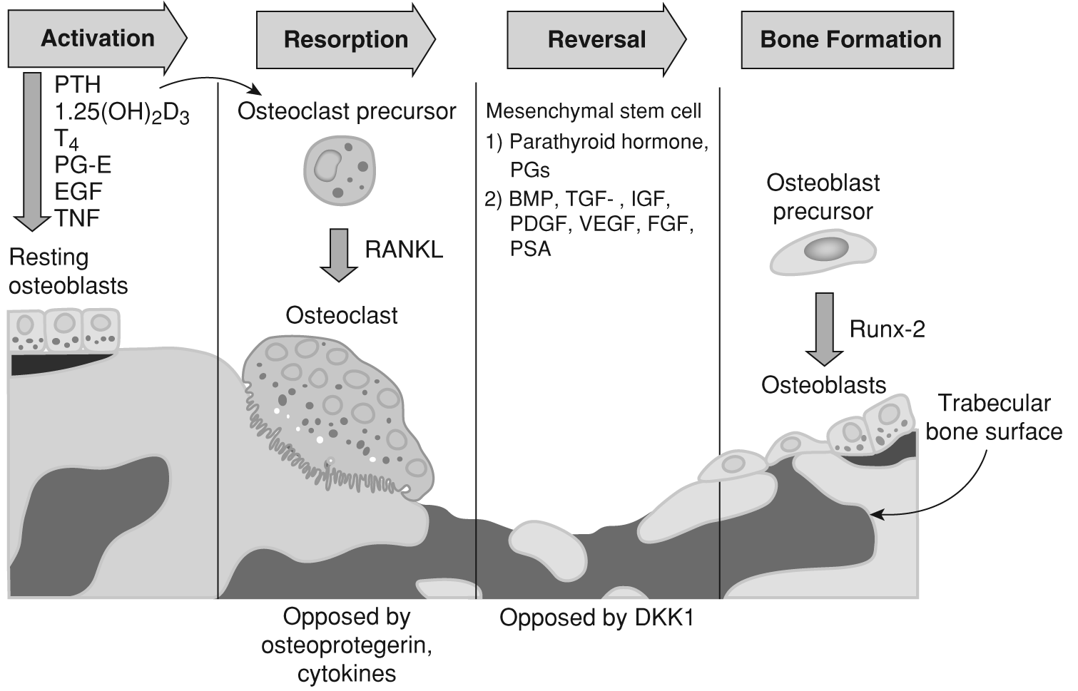
To review the basic physiology of bones, normal remodeling of the bone is constant during one's life, with osteoclasts breaking down bone cells (called resorption) and osteoblasts building new bone cell. To help keep these terms straight, it may help to think of the c in osteoclast as representing “catabolism” (meaning breakdown) and the b in osteoblast as representing “buildup.” Systemic as well as local factors control this process.
Osteoclasts are stimulated and regulated by systemic and local factors:
- Systemic factors: The hormones parathyroid hormone-related peptide (PTHrP), 1,25-dihydroxyvitamin D3, and thyroxine (T4) stimulate the process by inducing the marrow stromal cells and the osteoblasts to express receptor activator of nuclear factor-kB ligand (RANKL, also known as osteoclast differentiation factor).1 RANKL is also important in the immune system, as it helps dendritic cells to survive (survival factor) and helps to regulate thymus-dependent or T-lymphocyte–immune function. RANKL has been shown to have an important role in cell migration to the bone and in tissue-specific metastatic behavior of cancer cells.5
- Local factors: Osteoblasts secrete interleukin-6 (IL-6), IL-1, prostaglandin-Es, and colony-stimulating factors (CSFs), which lead to the formation of osteoclasts. Epidermal growth factor (EGF) and tumor necrosis factor (TNF) also stimulate osteoclastic activity, as can thymocyte-dependent lymphocytes (T-cells), which produce IL-17, thus stimulating the osteoclastic process.
Osteoclasts come from monocyte-macrophage precursors. Stromal cells in the bone microenvironment, as well as osteoblasts, produce macrophage CSF and RANKL. RANKL is released by activated T cells and is expressed on the surface of osteoblasts and stromal cells.1 The systemic factors increase (upregulate) the expression of RANKL on the marrow stromal cells and osteoblasts. RANKL initiates osteoclastogenesis and differentiation of osteoclasts by binding to the RANK receptor on the osteoclast precursors. Within the cell, the message gets sent to the nucleus of the osteoclast by the nuclear factor-kB (NF-kB) and Jun-N-terminal kinase (JNK) pathways. NF-kB is a transcription factor that turns on or off genes that control cell proliferation and survival. In many cancers, NFkB is dysregulated and it turns on unlimited tumor cell proliferation and survival. JNKs are members of the mitogen-activated protein kinase (MAPK) family and are responsible for interacting with stimuli from outside the cell (mitogens) and regulating gene expression as well as subsequent cell division, differentiation, proliferation, and survival or death (apoptosis). JNKs are also known as stressactivated protein kinases. Thus, as the message reaches the cell nucleus, it turns on the cellular genes to transform the cell into an osteoclast and to make sure the osteoclast survives.
The formation and activation of osteoclasts is tightly regulated. Local and systemic factors can halt the production of osteoclasts. Systemically, corticosteroids and T-cell–released IL-4, IL-18, and IF-gamma, together with locally produced TGF and osteoprotegerin (a cytokine called osteoclastogenesis inhibitory factor, a member of the TNF superfamily) inhibit the differentiation of macrophages into osteoclasts; osteoprotegerin works by binding to RANKL on osteoblast/stromal cells and osteoclast precursor cells.6 Thus, much like a scale, the balance or ratio of RANKL to osteoprotegerin regulates both the formation and activity of the osteoclasts.1
The osteoclasts act on the trabecular bone surface to break down the bone minerals and matrix. They release proteases (enzymes) that dissolve the matrix, and this process also releases an acid that removes the minerals,1 leaving small holes in the bone trabecula (framework of bone on the surface) where
the bone has been removed. Once they have done their job, the osteoclasts undergo programmed cell death (apoptosis).
The osteoblasts then come along and fill the holes with new bone that still must be mineralized or calcified. Mesenchymal stem cells produce the osteoblasts, and differentiation of the osteoblast into an adult osteoblast depends upon a transcription factor called Runx-2 (also known as core-binding factor- 1 [CBFA1]). CBFA1 turns on the known genes necessary for osteoblast differentiation.1 Bone morphogenetic proteins (BMPs) are growth factors and cytokines that stimulate the growth and development of osteoblasts. Systemic and local factors again control osteoblastic proliferation and differentiation.1 Factors that increase osteoblastic proliferation and differentiation are:
- Systemic factors: parathyroid hormone; prostaglandins; cytokines; and platelet-derived growth factor (PDGF)
- Bone matrix-released growth factors: bone morphogenetic proteins (BMPs); insulin-like growth factor (IGF); fibroblast growth factor (FGF); vascular endothelial growth factor (VEGF); prostate-specific antigen (PSA), which increases osteoblast proliferation; and endothelin-1, which increases osteoblast differentiation and proliferation.
Corticosteroids cause the osteoblast to undergo apoptosis; thus, they block new bone formation.1 The new bone has to be protected, so a lining of cells is laid over it. Old bone is replaced systematically so that building and breakdown are always balanced and the integrity of the human skeleton is intact (Figure 4-1). Normally, as women age, their bone buildup lags behind bone breakdown, and they are at risk for osteoporosis, a disease in which osteoclast action is greater than osteoblast activity. This risk intensifies as the woman reaches menopause at age 45–55 years7 and increases a woman's likelihood of osteoporosis and bone fracture. In hormonal breast cancer treatment with selective estrogen receptor modulators, women are at risk of losing bone density and are therefore advised to take calcium and vitamin D and to exercise regularly. Drugs like Fosamax slow the breakdown phase to help maintain bone mass.
The spectrum of bony metastases are anchored by osteolytic lesions on one end and osteoblastic lesions on the other, but in fact, patients may have both types. In general, patients with breast cancer have primarily osteoblastic lesions with about 15–20% osteolytic, while patients with prostate cancer have largely osteoblastic lesions.8 Patients with multiple myeloma have only osteoblastic bone lesions. Bone scans show the osteoblastic process (active bone formation).1
Process of Metastasis to Bone
Roodman describes a process that depends upon both tumor and microenvironment-produced molecules, highlighting the importance of the “seed and soil” analogy.1 The long bones containing red bone marrow receive high blood flow,
Figure 4-1. Process of bone building and breakdown
Data from Loberg RD, Logothetis CJ, Keller ET, Pienta KJ. Pathogenesis and treatment of prostate cancer bone metastases: Targeting the lethal phenotype. J Clin Oncol. 2005;23(32):8232-8241; Logothetis CJ, Lin SH. Osteoblasts in prostate cancer metastasis to bone. Nat Rev Cancer 2005;5(1):21-28; Roodman GD. Mechanisms of bone metastasis. N Engl J Med. 2004;350(16):1655-1664.
which makes it easy for embolized cells to find their way to bone. Breast, prostate, and myeloma tumor cells are able to make adhesion substances that allow the tumor cells to stick to the marrow stromal cells and surrounding matrix. Once the tumor cell adheres to the bone, the tumor cells ramp up the production of factors that turn up angiogenesis and bone resorption, as well as tumor proliferation.
Breast Cancer
Breast cancer cells can induce the osteoclasts to secrete PTHrP, TNF, IL-1, IL-6, IL-8, and IL-11, which in turn tell the osteoblasts to release RANKL.4 PTHrP and parathyroid hormone (PTH) both bind to the PTH receptor and effect the same physiologic results. PTHrP is produced by 92% of breast tumors metastatic to bone.9 The tumor cells also produce prostaglandin E2 and macrophage CSF.1 All these factors increase the expression of RANKL, which directly affects osteoclast precursors to make osteoclasts.
There are many inactive growth factors in the bone matrix that become activated by the process of bone resorption. Once the osteoclasts begin the bone resorption process, the destroyed matrix releases growth factors that in turn drive the tumor (growth) as well as turn up bone destruction. This process is me-
diated by the released growth factors from the destroyed matrix: BMPs, IGF-1, TGF, FGFs, and PDGF. The stimulated breast cancer cells release more PTHrP, and the vicious cycle is heightened.10
Of course, calcium is released when the bone matrix is destroyed, thus raising the patient's serum calcium. Elevated calcium levels in turn stimulate breast cancer cell growth as well as the production of PTHrP. This occurs because there is an extracellular calcium-sensing receptor (CaR) that normally senses elevated calcium levels in the parathyroid and kidney, which then respond to correct the imbalance. However, in cancers of the breast, it appears the CaR actually stimulates the release of PTHrP from the tumor, along with the other osteoclastdriving growth factors, which drives further bone resorption and further release of calcium, resulting in the well-known hypercalcemia of malignancy.11
Additionally, it appears that breast cancer cells that produce osteolytic lesions in bone have increased levels of Wnt/beta-catenin signaling and expression of dickkopf1 (DKK1) (an antagonist to Wnt/beta-catenin that also inhibits osteoblast differentiation and osteoprotegerin expression, thus increasing osteoclast activity). Bu et al. suggest that breast-cancer–produced Dkk1 is an important link between primary breast cancers and secondary osteolytic bone metastases.12 Although most breast cancer bony metastases are osteolytic lesions, some are also osteoblastic, or both. Again, both systemic and local factors are released, driving the formation of unstable bone. Systemic factors are PTH, prostaglandins, and cytokines as well as lymphocyte-produced PDGF, which together with the locally (bone matrix) produced BMPs, TGF, IGFs, and FGFs, drive the osteoblastic process.1 It is thought that osteoblastic lesions occur as a result of endothelin-1 stimulation. Similarly, there is a vicious cycle: As the tumor cells release or induce endothelin-a, osteoblastic bone remodeling forms unstable bone, and the osteoblasts release growth factors such as more endothelin-1, PDGF, and urokinase (made by osteoblasts in the bone microenvironment). This then stimulates breast cancer cell growth and release of systemic factors, which together with local factors, drive the osteoblastic cycle around again.
Prostate Cancer
Prostate cancer bony metastases are primarily osteoblastic. Prostate cancer cells secrete osteoblast-stimulating factors (e.g., PDGF, IGFs, Wnt family ligands that turn on the signaling pathway for osteoblastic activity, endothelin-1, BMPs, urokinase-type of plasminogen activator.1 4 PSA is able to break (cleave) the PTHrP molecule, so this may be a way osteoclastic activity is turned off. In addition, it appears to activate osteoblastic growth factors that are released from the bone matrix during bone building (osteoblastic activity): IGF-1, IGF-2, and TGF. However, although there is no evidence of increased osteoclast activity pathologically in the bone, it appears that bone resorption does occur, either before osteoblastic activity or after, as bone-resorptive (osteoclastic)
markers are increased in patients with bony metastases (serum C-terminal telopeptide of type I collagen and tartrate-resistant acid phosphatase), and it appears that blocking osteoclastic activity decreases skeletal events in patients with metastatic prostate cancer with bony metastases.1 13 14
Multiple Myeloma
In multiple myeloma, osteoclasts accumulate next to myeloma cells on bone-resorbing surfaces, and it is the osteoclasts, not the tumor cells, that destroy the bone matrix.1 It appears that IL-1, IL-6, macrophage inflammatory protein-1 (MIP-1, a chemotactic cytokine also called CCL3), and RANKL are the culprits in ramping up the osteoclasts.1 RANKL is produced in large measure by the myeloma cells. MIP-1 is produced by activated macrophages and induces the synthesis and release of the proinflammatory cytokines IL-1, IL-6, and TNF. It is elevated in about 70% of patients with multiple myeloma, and it is not only important in increasing bone destruction and myeloma cell survival, but also enhancing the ability of myeloma cells to stick to the bone surface.15 It does this by upregulating the myeloma cells' expression of 1 integrins (“grappling hooks”) so that the cell can adhere to the bone surface; once this happens, the communication between the marrow stromal and myeloma cells amplifies the production of IL-6, RANKL, and MIP-1.1 Of course, this increases the vicious cycle of bone breakdown.
Just how osteoblast formation and differentiation is inhibited is less clear and is probably multifactorial. TNF released by the myeloma cells can prevent growth and differentiation of osteoblasts, as can DKK1, which is expressed on myeloma cells.1 DKK1 is an antagonist of the Wnt-signaling pathway, which is the principal pathway that brings the message to the cell nucleus to activate the genes that will move the precursor cell into an osteoblast. Giuliani et al. showed that myeloma cells block RUNX2/CBFA1 activity in the bone marrow osteoblast progenitor cells, thus inhibiting osteoblast formation and differentiation.16 Because bone scans can pick up only osteoblastic density, and because there is none in multiple myeloma (osteolytic without any osteoblastic response), the bone scan may appear normal despite the presence of osteolytic lesions.1
Targets include:
- RANKL, by increasing the level of osteoprotegerin with recombinant osteoprotegerin to turn off or downregulate RANKL, or to block RANKL by a monoclonal antibody (MAb) targeted against RANKL
- Prevention of the osteoclast from adhering to the bone surface so it is not able to perform bone resorption, or inhibition of the proteases released by the osteoclast so it cannot make holes in the bone (e.g., cathepsin K)
- RUNX2/CBFA1 to prevent the block by myeloma cells so that osteoblast formation and differentiation can occur
- Endothelial-1 (ET-1), as it activates the ETA-receptor, which facilitates prostate cancer progression (proliferation, avoidance of apoptosis, invasion) as well as new bony metastases; ET-1 also mediates the interaction of prostate tumor cell with bone microenvironment (osteoblast, osteoclast, stroma)17
Currently, two bisphosphate drugs are approved by the Food and Drug Administration (FDA) for the treatment of malignant hypercalcemia and the treatment of patients with bony metastases. The first approved was pamidronate (Aredia) for the treatment of patients with osteolytic lesions related to either breast cancer metastases or multiple myeloma. The second was zoledronic acid (Zometa), which is indicated for the treatment of patients with multiple myeloma; patients with documented bone metastases from solid tumors, in conjunction with standard antineoplastic therapy; and patients with prostate cancer that has progressed after treatment with at least one hormonal therapy. There is evidence that women treated with these agents have decreased skeletal events or increased disease-free survival.18 20
Further exploration into the mechanism of bone metastasis has revealed a dramatic story involving RANKL. As discussed, RANKL activates bone osteoclasts so that they start to break down bone. In cancer, there is a vicious circle of tumor-released factors that stimulate osteoclast activity, mediated by RANKL; breakdown of the bone matrix releases growth factors that stimulate both tumor growth and continued bone breakdown. Amgen has submitted an application to the FDA for consideration of denosumab (Prolia), an investigational RANKL inhibitor, for the treatment and prevention of bone loss in patients undergoing hormone ablation therapy for either prostate or breast cancer, and for treatment and prevention of postmenopausal osteoporosis in women.21 Denosumab, is a fully human MAb that targets RANKL, by mimicking osteoprotegerin, which is a soluble decoy receptor for RANKL that blocks ligand binding to RANKL so it does not become activated and the message (signaling) is not sent to start differentiation and activation of the osteoclast.5 22 In addition to being critical for osteoclast differentiation, function, and survival, when RANKL is released in large amounts from the bone marrow, it attracts cancer cells that express its receptor, RANK, to the bone where they can develop a metastatic site, and the microniche is well prepared to encourage strong metastatic growth.5 23
Denosumab has shown to be superior to the existing bisphosphonate drugs. In a large (N = 2049) international phase III, randomized, double-blind clinical trial comparing denosumab with zoledronic acid in the treatment of bone metastases in patients with advanced breast cancer, the group receiving denosumab had significantly longer time to first on-study skeletal-related events (SRE, e.g., fracture, radiation to bone, surgery to bone, or spinal cord compression [hazard ratio 0.82]) and longer time to the first and subsequent SREs (hazard ratio 0.77). The primary end point was to evaluate if denosumab was noninferior to
zoledronic acid in the prevention of first on-study SRE, and the second end point was to see if denosumab was superior to zoledronic acid in terms of first on-study SRE as well as subsequent SREs. Both end points were met.24 In prostate cancer, denosumab was effective in reducing N-telopeptide urine levels (marker of osteoclast activity) in patients who previously had received intravenous (IV) bisphosphonates.24 25 Prostate cancer patients represented 45% of the total; the remainder was made up of patients with bony metastasis from other solid tumors and multiple myeloma. At week 25, 69% of the denosumabtreated patients continued to have urine N-telopeptide levels < 50 compared to 31% of the patients treated with IV bisphosphonates.
In addition to therapeutic benefit in reducing skeletal events from bony metastases, denosumab offers benefit to the many women and men who develop bone loss from endocrine therapy. Women with breast cancer receive aromatase inhibitors for 5 years. Although this treatment is intended to block estrogen effects, it also enhances and accelerates bone loss. While treatment with tamoxifen has been shown to benefit bone, the aromatase inhibitors, such as the third-generation anastrozole, letrozole, and exemestane, increase bone loss beyond that seen naturally after age 30 years.26 Denosumab offers a benefit to these patients as well. Ellis et al., in the phase III HALT breast cancer study, showed that denosumab 60 mg administered every 6 months subcutaneously to women receiving adjuvant aromatase inhibitor therapy (N=252) increased bone mineral density (BMD) in the lumbar region by an average 5.5% over that of patients receiving placebo at 12 months; at 24 months, BMD increased an average of 7.6% over placebo. Increases were also seen in total hip (4.7%), femoral neck (3.5%), trochanter (5.9%), and radius (6.1%).27 Increases in lumbar spine BMD were apparent within 1 month of therapy and were not influenced by length of time on aromatase inhibitor therapy. The overall adverse effects were similar to placebo. Similarly, the phase III HALT prostate cancer study (N=1400) of men with prostate cancer undergoing androgen deprivation therapy (ADT) met both its first and second end points by demonstrating significant increases in BMD in the lumbar spine compared to placebo at 2 years and also showing half the incidence of SREs (e.g., fractures) compared to placebo at 3 years, respectively.28 29 Men with prostate cancer who survive at least 5 years with prostate cancer and have received ADT are estimated to have a 19.4% incidence of bone fracture, compared to 12.6% for those who did not receive ADT.30