There are many differences between normal and malignant cells.
Normal cells look like the parent cell, with a distinct shape and appearance. The nucleus is small, and the nuclear to cytoplasmic ratio is small, as opposed to malignant cells where the ratio is high. The normal cell is differentiated with a clear function(s); for example, erythrocytes make hemoglobin. Normal cells bind closely together and stay put in their neighborhood. They stop dividing when they feel their neighbors (contact inhibition) and are not able to move from their neighborhood (anchorage dependency). Normal cells have a diploid number of chromosomes (23 pairs, or 46 individual chromosomes), which is called euploidy. In contrast, cancer cells may have fewer or more copies of the chromosome, called aneuploidy.
As stated previously, normal cells divide only when the body needs more cells. Once an individual is born, the body is very careful that cell birth equals death, so that cell division occurs only when needed. Proto-oncogenes are genes that drive cell division. One can think of them like the accelerator of a car, pushing the cell to divide when the body communicates the need. The safeguards of this are TSGs, which can be thought of as the brakes on cell division, so that when cell birth and death are equal, the TSGs stop the proto-oncogenes from pushing for cell division. Because millions of cells need to be replaced daily, many mistakes (mutations) can occur in the replication of the cells. DNA repair genes either excise or repair these mutations or mistakes in copies of the DNA when the cell is dividing; if unable to repair them, the cell is sent to programmed cell death (apoptosis). In cancer, there appear to be mutations in proto-oncogenes, forming oncogenes, TSGs, and the DNA repair genes. In this way, the accelerator on cell division becomes unopposed.
Let's get back to normal cell division. Mitosis is the division of the cell into two daughter cells. The normal cell cycle is tightly controlled, as has been briefly discussed. There are restriction points that allow the cell to check whether the cell has the right amount of DNA, whether it is correctly copied, and whether it divides properly. The cell cycle is controlled by proteins made by TSGs, and, unfortunately, many of them are mutated or silenced by cancer.
Not all cells are dividing. Some remain in the resting or G0 phase of the cell cycle. When cells are in the G0 phase, they are actually busy doing their daily jobs as differentiated cells. If the body says it needs more cells, then cells are recruited from G0 to enter the cell cycle. For example, during the digestion of food, the microvilli in the intestinal crypts shed about 80 million cells per minute.5 The body needs to replace these cells. That is why these cells are a highly proliferating normal cell population.
The term cell cycle describes the journey of a cell to replicate or copy itself, resulting in two identical daughter cells. Early scientists knew what happened in the
synthesis (S) phase and the mitosis (M) phase, but they did not know what happened in the periods between these phases, so they called them gaps (hence the G). G1 is the time before the S phase, and G2 is the time after the S phase and before the M phase. Cyclins, which are subunits of CDKs, regulate the cell's journey through the cell cycle. The movement of the cell through each phase of the cell cycle depends upon the synthesis of the correct cyclin. The cyclin binds to its CDK, and together they form an enzyme that activates other proteins via phosphorylation. While the CDK level stays about the same as the cell goes through the cell cycle, the cyclin level rises and falls, which is how the term cyclin was derived. The cyclins are degraded after mitosis so that the cell cycle can be shut off as a safety mechanism. When control of the cell cycle goes awry, as in cancer, chromosomes with mutations or damage can continue to go through the cell cycle, resulting in genetic instability. For example, normally the cell will divide into two daughter cells, each with 23 pairs of chromosomes. When the checkpoints of the cell cycle are ignored, as with cancer, cells can end up with fewer or more chromosomes (aneuploidy), or with gene rearrangement with parts of one chromosome ending up on another chromosome (translocation). Probably all cancer cells have mutations in the checkpoint genes and in the genes that control cell cycle progression.
G0 Phase and Entrance into G1
Once a cell is recruited into the cell cycle, it needs to make sure that the body can support new cells. The cell advances to the G1 restriction point, and the cell then assesses if indeed the body needs more cells, if there is adequate nutrition to support the new cells (e.g., oxygen, glucose), if the cell is big enough, and if it has the resources to complete the replication (e.g., energy, proteins to make the membranes, organelles, DNA). If the answer is yes, then the cell proceeds to the S phase. This decision is irreversible once the cell begins to cycle. If the answer is no, the cell goes back into G0.
The gene product is the protein that the gene codes for, and it is actually the protein that does the work of the gene. The protein product of the Rb TSG, pRb, regulates G1 progression. In early G1, pRb is in a hypophosphorylated state. It binds tightly to and represses (shuts off) the activity of the E2F family of transcription factors that need to be available to make the right genes expressed (turned on) when the cell gets to the S phase.
When the cell leaves G0 and goes into G1, CDK4 and CDK6 actively combine with D-cyclins (D1, D2, D3), which begin phosphorylation of the pRb; this lifts the hold on the E2F transcription factors so they can get the genes ready in the S phase. This process is critical for the cell to pass the restriction point.
A final check is made to see if there is any damage to the DNA; if so, the cell is not allowed to enter the S phase. If DNA damage has occurred, p53 accumulates in the cell, induces p21-mediated inhibition of cyclin D/CDK. When cyclin D/CDK complex is inhibited, the pRb is in a state of low phosphorylation and is tightly bound to the transcription factor E2F, which inhibits Rb.
Entrance into the S Phase
Once the cell gets the OK to pass into the S phase, the rest of the cell cycle is well programmed. Cyclin E picks up from cyclin D when pRb interacts with histone deacetylase proteins (HDACs), which are involved with chromatin remodeling.6 Now that cyclin E is expressed, it complexes with CDK2 (CDK/cyclin E), keeping the pRb phosphorylated so the cell cycle can continue to progress. Next, cyclin A enters the picture and becomes the primary partner of CDK2 (CDK2/cyclin A). In order to set up replication of the chromosomes during the S phase, CDK2, which is now released from its cyclin, targets a protein Cdc6, which is necessary to start replication. It becomes phosphorylated (turned on) by CDK2/cyclin A and moves to the cytoplasm where it complexes with E2F.
The cell now replicates the chromosomes, and like a detective with a magnifying glass, the DNA damage checkpoint inspects the DNA to see if it is damaged. If so, progress is halted until it can be repaired.
Entrance into G2
Cyclin A continues to G2 but changes partners, now complexing with Cdc2 (cell division cycle 2, also known as CDK1).
The cell goes through the G2 checkpoint; if DNA replication is not completed, the cell cannot enter the M phase.
If the replicated DNA is complete and there are no mistakes, then the cell is ready to begin mitosis. To pass through, cyclin B now replaces cyclin A and complexes with Cdc2.
Entrance into the M Phase
The cyclin B/Cdc2 complex phosphorylates proteins regulated during mitosis.6
Aurora kinases are very important enzymes that control chromatid segregation and make sure that the genetic material is correctly shared in each daughter cell. Aurora A functions during the first phase of mitosis (prophase), ensuring that the centrosomes make the mitotic spindle upon which the chromosomes line up. Aurora B helps the mitotic spindle attach to the centromere (the condensed and constricted area of a chromosome, like the waist, to which the spindle fiber is attached during mitosis). Aurora C's function is poorly understood. All or some may be mutated in cancer. See Chapter 4 for a discussion of aurora kinases as molecular targets.
Polo-like kinases (Plk1s) also are very active during the M phase. Plk1s help ensure the mitotic spindle is put together correctly and also can activate or inactivate CDK/cyclin complexes. They may be mutated in cancer.
The cell is checked to make sure the chromatids are assembled properly on the mitotic spindle, and if not, the cell cannot enter anaphase.
One group of proteins that can inhibit the CDKs by binding to them is the INK family (inhibitors of CDK4). This family includes p164, which inhibits CDK4,
and three others that bind to and inhibit CDK4 and CDK6: p154, p184, and p194. These TSGs are often mutated in cancer, which allows the imperfect cell to continue through the cell cycle. Another family of proteins called CDK interacting protein/kinase inhibitory protein can bind to and inhibit many CDKs, but also may promote the activity of cyclin D-dependent kinases. Many tumors have subverted these inhibitory signals so that the progression through the cell cycle is not halted.6 8
Because the cell cycle is almost universally flawed in cancer, it presents many possible targets, and CDK inhibitors, DNA damage checkpoint kinase inhibitors, aurora kinase inhibitors, and Plk inhibitors are all being studied.7
The Multistage Model of Carcinogenesis
Cancer is a disease of mutations, and it is estimated that malignant transformation requires at least four separate mutations in key genes. Approximately 300 genes have been found mutated in cancer.7 Malignant tumors are clonal; a group of cells with invasive and aggressive features are self-selected and then become invasive and metastasize. However, there is mounting evidence that cancer stem cells may play a key role in driving invasion and metastasis. Key genes that are mutated in cancer are proto-oncogenes, which then form oncogenes, TSGs, and DNA repair genes. In addition, often there are mutations in genes controlling the signaling cascade that can be turned on so that the message to divide and invade is continually sent. There is also increasing awareness that the tumor microenvironment ensures that the tumor has the necessary signals to grow, invade, and metastasize.
To review, normal cells obey very strict rules about how they behave, and they divide only when the body needs more cells. They respond to contact inhibition, so that the pressure of neighboring cells negates any stimulus to divide. It is almost as if the cells have an “area code” within which they must stay; if they leave the area, they will undergo programmed cell death or apoptosis. An exception to this is the neutrophil, which is able to migrate to areas of infection. Otherwise, normal cells stay where they are supposed to stay, grow only in one layer, and stop dividing when they touch their neighbors. Normally, when the cells do divide, they do it faithfully. If there are mutations in the DNA of the new cell, the DNA repair gene products try to excise or repair the mistake; if repair or excision is not possible, the cell is sent to programmed cell death, directed by P53 gene and its protein product p53.
If there are mutations making this usual process ineffective, then cells with mutated genes accumulate and continue to divide, making more and more mutations. As billions of cells in the body are replaced every day, mistakes do occur. If the mutation is on or near a proto-oncogene, or a TSG, then the stage may be set for malignant transformation.
There are many mutations in DNA because so many cells are replaced daily. These mutations can be subtle alterations in the spelling of a gene, so that an
incorrect protein is produced by the gene. This mutation can be harmless and is usually repaired by DNA repair genes. If the mutation is on or near a protooncogene, an oncogene can result, which can drive cell proliferation, new blood vessel formation, and other factors necessary for invasion and metastasis. Genetic mutations seen in cancer include subtle alterations such as small letter deletions, insertions, and single base pair substitution, such as a point mutation. They can involve:
- Chromosome number changes such as aneuploidy or loss of heterozygosity (LOH; loss of normal function from one copy of the gene or allele when the other copy is already inactive)
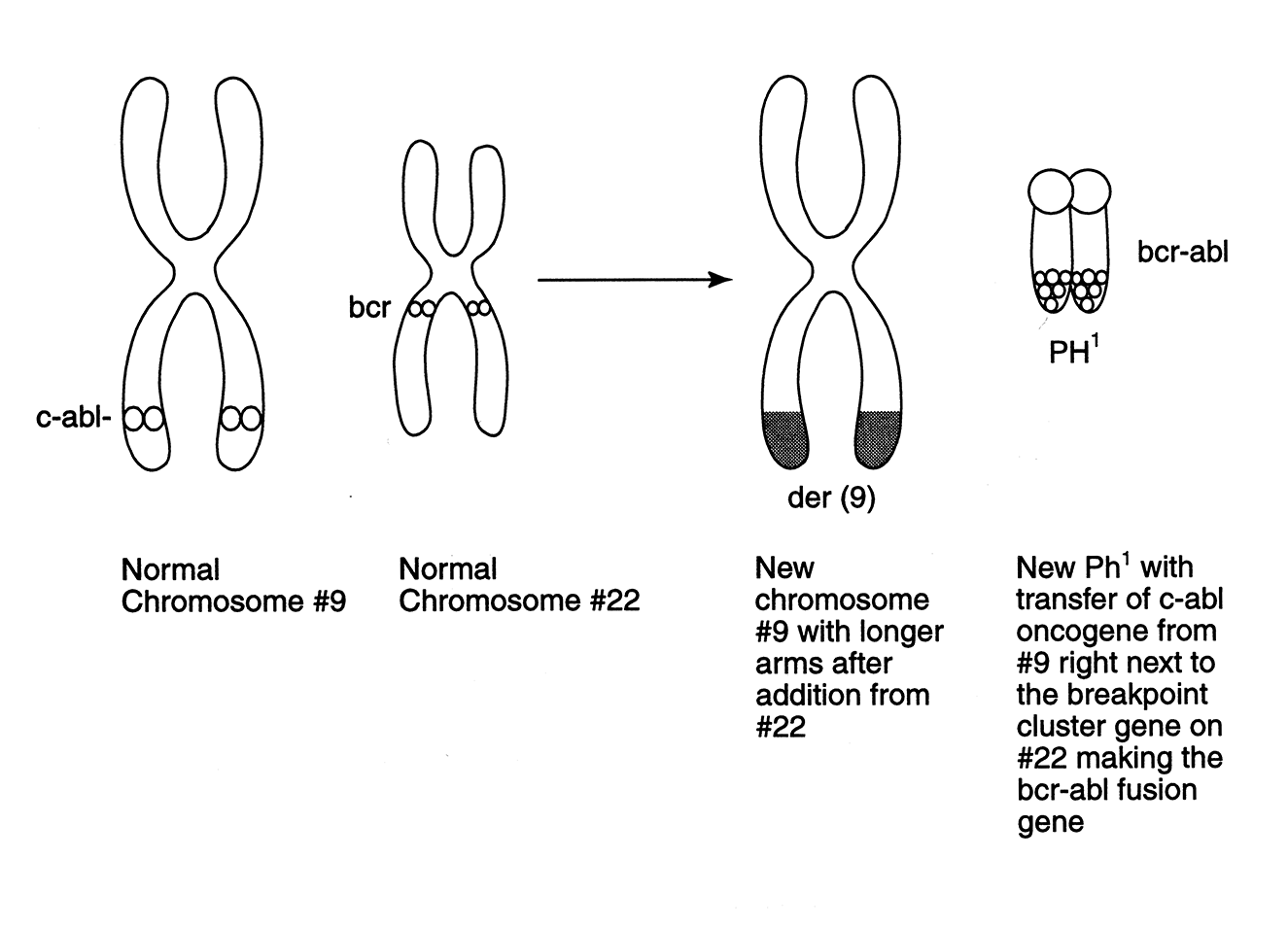
- Chromosome translocation wherein part of one chromosome ends up on another chromosome (if it lands near or on a proto-oncogene, it can result in an oncogene, as seen in Ph1-positive chronic myelogenous leukemia [CML], depicted in Figure 1-1)
- Amplification of oncogenes (such as with HER2-positive breast cancer, where there are 5- to 100-fold more copies of a gene in a small region of a chromosome), or an amplicon that contains one or more genes that increase cell proliferation
- Exogenous sequences such as a virus encoded in a cell's DNA that drives malignancy, such as HPV in cervical cancer, Epstein–Barr virus in Burkitt lymphoma, and hepatitis B, which leads to hepatocellular carcinoma
In cancer, there are mutations in the key genes that control the normal cell division scenario. A person has two copies of each gene, one from the mother and one from the father. People with a hereditary predisposition to cancer, such as familial polyposis coloni, inherit one copy of the damaged gene, but the other copy is usually normal. These people are considered heterozygous for the trait the gene encodes. Gene mutations that are inherited occur in the germ line; thus all the person's cells have this mutation. Through LOH, the normal gene mutates and the person loses its function so that now both genes are abnormal. Individuals without an inherited predisposition develop acquired mutations in somatic cells (i.e., the mutation occurs in regular cells, not germ line cells and is therefore not in all cells and cannot be inherited). In the course of these individuals' lives, first one, then the second copy is mutated, and the journey to malignancy begins.
Most mutations are not inherited. They occur through daily life, such as exposure to sun (ultraviolet light), the foods we eat, smoking (if we smoke), and aging, as well as through the wear and tear of normal cellular processes.
Proto-oncogenes drive cell division when the body needs cells. In malignancy, there are mutations in the proto-oncogene, forming an oncogene, which continues to send messages to the cell nucleus to divide, much like putting one's foot on a car's accelerator. Oncogenes are activated, or “turned on,” so that they continue to send the message to the cell nucleus to divide, as if the accelerator pedal were pressed to the floor. When the oncogene is mutated, it matters where on the chromosome the oncogene is positioned. Table 1-1 shows some examples of oncogenes. There are at least 100 oncogenes known at this time, and they fall within five major classes10:
Figure 1-1. Philadelphia chromosome: example of a reciprocal translocation and formation of an oncogene
Wilkes and Barton Burke.9 Reproduced with permission from the publisher.
- Growth factors—These factors stimulate cell growth (e.g., c-sis overexpression results in the overproduction of PDGF).
- Growth factor receptors, such as the EGFR1 (erbB1) and EGFR2 (HER2 or erbB2)—The receptor should be turned on when a growth factor binds to it, but when the gene is mutated, the receptor can be turned on indefinitely, constantly sending the message for cell division to the cell nucleus. The genes erbB1 and erbB2 can also be amplified, which means the cell has too many copies of the gene rather than the normal two copies, again resulting in many signals being sent to the nucleus of the cell for the cell to divide.
- Signal transducers, such as ras and abl—These proteins help pass the message along to the cell nucleus. They are usually turned on or off based on the cell's need for signaling. When mutated, they stay on and constantly send messages to the cell nucleus to make the cell divide.
- Transcription factors, such as myc—These factors are at the DNA level and direct the copying of the genes (recipes for protein[s]), which tell the cell to divide or other functions. They are normally turned on when needed, and off after they copy the gene's recipe for the protein so that no more proteins will be made. When mutated, these transcription factors can remain on. Some transcription factors are activated when mutated in many cancers, and initiate and maintain the tumor phenotype (behavior) in remodeling the tumor microenvironment with inflammatory and other mediators.4
| Oncogene | Function | Comments/Cancer Types |
|---|---|---|
| v-sis | Overexpresses and produces platelet-derived growth factor | Located on long arm chromosome 22; prostate |
| Erb1 (EGFR) | Increased cell proliferation, survival, angiogenesis, invasion, and metastases | Can be mutated or amplified; CRC, breast, lung, SCHNC, pancreatic |
| Erb2 (HER2) | Increased cell proliferation | Increased signaling or amplification; breast, gastric, lung |
| Ras | Increased cell proliferation | Pancreatic; K-ras colon; H-ras breast, melanoma, lung, kidney, bladder; NRAS ovarian, thyroid, melanoma, leukemia |
| Abl | Increased cell proliferation | CML |
| Myc | Increased cell proliferation, malignant transformation, remodeling of stroma | C-myc Burkitt lymphoma; N-myc neuroblastoma |
| Bcl2 | Prevents apoptosis of abnormal cells | Lymphoma |
| BRAF | Involved in cell signaling; helps to regulate the MAPK signaling pathway, which is very important in cell growth and differentiation | Gastric, colon |
| RET | Its protein is a receptor tyrosine kinase that is important in communicating messages from outside the cell. It is essential in the development of normal kidneys and the nervous system. RET stands for “rearranged during transfection.” | Thyroid, multiple endocrine neoplasia |
| MET | Makes receptor for hepatocyte growth factor (HGF)/scatter factor; important regulator of cell proliferation, motility, invation, and survival | Papillary renal cell cancer |
- Regulators of apoptosis (programmed cell death), such as the gene bcl-2— When mutated, the gene becomes hyperactive and dominant, and prevents programmed cell death of abnormal cells.
TSGs halt cell growth when the body has the exact number of cells needed, again so cell birth equals cell death, and work much like the brakes on a car. In cancer, the TSGs are damaged, either through inactivation or through loss of the part of the chromosome that contains the TSG (locus). Unlike oncogenes, which are turned on and continue to send signals for growth, TSGs are turned off or inactivated so that they do not work. With first one copy of the TSG mutated, the brakes on cell division begin to fail, and when the second copy of the TSG is mutated or lost, the brakes really fail, so that cell division cannot be opposed. Table 1-2 shows examples of TSGs. Types of TSGs can be classified in terms of their function10:
- Genes that control cell division, such as the retinoblastoma gene (Rb1)— These genes are crucial for guarding the cell cycle from incompetent cells that should not pass through the cell cycle.
- Genes that repair DNA—These genes are crucial for “proofreading” the DNA strands after they have been replicated to identify any mistakes and to fix them by either excising the mistakes or correcting the misspelling (base pairs) in the DNA strand. People with hereditary nonpolyposis colon cancer (HNPCC) are born with one or more DNA repair genes that are mutated and cannot correct the errors in DNA.
- Genes responsible for apoptosis, such as p53—These genes are the guardians of the genome and the gene product that forces the irreparably mutated cell into programmed cell death (see Chapter 4). Many solid tumors have mutations in p53. Individuals with the Li-Fraumeni syndrome inherit p53 mutations and have a high risk for developing multiple cancers.
DNA is the building block of life and contains a person's complete genetic information (the “blueprint” for that individual), such as eye and hair color, as well as all the instructions to operate the billions of cells in the body. DNA is highly protected and is the only biomolecule in the body that is repaired, with all other defective biomolecules being replaced.11 There are four general systems of DNA repair: (1) direct damage repair, (2) excision of the damaged DNA portion, with precise replacement of the correct sequence of nucleotides, (3) double-strand break repair, and (4) damage bypass. The excision of a damaged DNA portion and replacement of correct sequence can be subdivided into three types: (1) mismatch repair, (2) base excision repair, and (3) nucleotide excision repair.
| Tumor Suppressor Gene | Function | Comments/Cancer Types |
|---|---|---|
| TP53 | Transcription factor responsible for detecting irreparable DNA mutations and causing them to undergo apoptosis | Mutated in many solid tumors |
| RB1 (retinoblastoma) | Normally guardian of the genome; when mutated, the cell cycle is uncontrolled | Retinoblastoma, cervical |
| DNA Repair Genes MLH1, MSH2, or MSH6 | Repair of DNA mutations | Hereditary nonpolyposis colon cancer (HNPCC) |
| BRCA1, BRCA2 | DNA repair genes, act as tumor suppressor genes | Inherited susceptibility to breast (BRCA2), ovarian, or pancreatic |
| APC | Makes a protein that communicates with other proteins to control whether cell division occurs and to ensure that the correct number of chromosomes is present in the cell during cell division; helps verify adherence to neighboring cells and cell migration | CRC, gastric, pancreatic |
| ATM | Controls the rate of cell growth and coordinates the repair of damaged DNA by working with BRCA1 and other DNA repair genes | Leukemia, lymphoma, breast, ovarian |
| CDK4 | Cyclin-dependent kinase that, together with CDK2, determines whether the cell will continue in cell division at the G1/S control point in the cell cycle | Melanoma |
| DCC | Helps regulate cell death and is important in axon guidance | Colorectal |
| INK4A | Codes for 2 proteins (p), which help to control the cell cycle: p16 stops the cell cycle in the G1 phase, and p19ARF stops p53 from being broken down | Melanoma |
| PTEN | Codes for the PTEN protein, which helps to regulate the cell cycle, thus preventing rapid cell growth and division | Breast, prostate, endometrial |
| VHL | Helps regulate the cell cycle, preventing rapid cell growth and division; works with other proteins to break down proteins when no longer needed, such as hypoxia-inducible factor (HIF), a key player in angiogenesis | Renal cell, pheochromocytoma, hemagioblastoma |
In cancer, unfortunately, often the DNA repair genes are mutated so that cells with significant mutations can progress through the cell cycle and reproduce cells containing mutations. With each division, more mutated cells go through cell division, so that more cells with additional mutations are produced. Eventually a clone of cells with particularly aggressive behavior (phenotype) will be selected (selection of the fittest).
As mentioned, hereditary mutations in DNA repair genes often lead to malignancy, such as HNPCC, which is associated with mutations in the DNA mismatch repair (MMR) genes. The mutations prevent the gene from giving the correct recipe for making the repair protein. When MMR genes are unable to fix “spelling” errors in the DNA before mitosis, the spelling errors or “mismatches” accumulate, possibly leading to oncogenes or damaged TSGs.
If a person has a mutation in both copies of the MMR genes, such as mutations in both copies of the MSH2 or MLH1 gene (homozygous), then the person is at risk for not only developing colon and other cancer but also for leukemia, lymphoma, or neurofibromatosis (called CoLoN, for Colon tumors, and/or Leukemia, and/or Neuroblastomatosis) at an early age.12
Most patients (90%) with HNPCC colon cancers show a phenomenon called microsatellite instability (MSI), compared to only 15–20% of patients with sporadic colon cancers. Microsatellites are short, repeated sequences of DNA. Recall that
the DNA sequences are made up of nucleotides that are represented by the four letters in DNA base pairing, A (adenine), T (thiamine), C (cytosine), G (guanine), where A always is matched with T, and C with G. The number of repeats at a single point in the DNA helix may differ among people in a given population, but in each individual, it has a constant length. For example, at a single place in the DNA strand (locus), one might have three AC repeats and another population may have four AC repeats at that same locus. But when an individual is born, he or she has a constant number of DNA repeats or microsatellites. Over time, there are mutations; this number can increase and sequences can be abnormally long or short.
In cancer, if there are more or less repeats of these DNA sequences in the microsatellite regions of DNA than in the DNA the person was born with, then MSI is indicated. When a laboratory does a test for MSI, the lab examines five different microsatellite regions of DNA. MSI-H(igh) is usually considered if microsatellite changes are found in two or more microsatellite regions; MSI-L(ow) is considered if changes are found in only one region, and MSS (stable) is considered if no changes are found. MSI-H is often the result of mutations in the DNA repair genes so that the mutations cannot be repaired. In HNPCC, MSI-H patients have a better prognosis than patients with sporadic colon cancer that is either MSS or MSI-L.13 Additionally, there is evidence that 5-FU–based chemotherapy confers no benefit in patients with MSI-H colon cancer and may be harmful.14
Other well-known DNA repair genes that are mutated in cancer and that become targets for therapeutic intervention include BRCA1 and BRCA2, which are associated with the homologous recombination repair pathway and an increased risk for developing breast cancer. Both are TSGs. BRCA2 works to repair DNA double-strand breaks together with a molecule called RAD51. BRCA1 helps to repair DNA errors by interacting with other members of the DNA repair system and regulating the expression (turning on or off) of genes that are involved in the repair of DNA mistakes. If BRCA1 and/or BRCA2 are not functioning, then cells with abnormal chromosomes accumulate and continue to mutate, and malignant transformation can occur.15 If BRCA1 and BRCA2 are not functioning, then a backup DNA repair process involves poly (ADP-ribose) polymerase (PARP) enzymes. PARP enzymes are needed to repair DNA following damage from internal or external stressors. PARP inhibitors selectively kill breast cancer cells with defective BRCA1 and BRCA2 repair genes. Normal cells with functional BRCA1 and BRCA2 are able to use a secondary, backup repair process to repair DNA damage, while BRCA1 and BRCA2 mutated malignant cells now have both pathways blocked.16 See Chapter 4 for a discussion of PARP1 inhibitors.
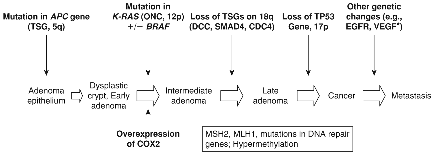
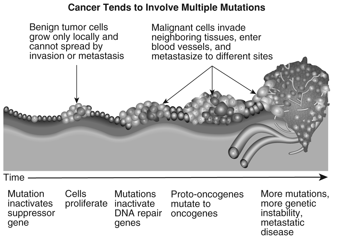
It is known now that there are many different pathways to malignancy even within a tumor type, such as that observed in breast cancer and colon cancer. See Figure 1-2 for a sequence of mutations leading to malignant transformation in colon cancer. Figure 1-3 shows the development of metaplasia, dysplasia, carcinoma in situ, and finally invasive cancer and metastasis.
Figure 1-2. Mutations in colorectal cancer (CRC)
Data from Deng Y, Kurland BF, Wang J, et al. High epidermal growth factor receptor expression in metastatic colorectal cancer lymph nodes may be more progrnostic of poor survival than in primary tumor. Am J Clin Oncol. 2009;32(3):245-252; Fearon ER, Vogelstein B. A genetic model for colorectal tumorigenesis. Cell 1990;61:759–767; Hisamuddin IM, Yang VW. Molecular genetics of colorectal cancer: An overview. Curr Colorectal Cancer Reports 2006;2(2):53-59; Kabbinavar F, Hurwitz HI, Fehrenbacher L, et al. Phase II, randomized trial comparing bevacizumab plus fluorouracil (FU)/leucovorin (LV) with FU/LV alone in patients with metastatic colorectal cancer. J Clin Oncol. 2003;21:60-65; Walther A, Johnstone E, Swanton C, et al. Genetic prognostic and predictive markers in colorectal cancer. Nature Rev Cancer 2009;9:489-499.
While a number of independent, different pathways are being identified, the following diagram highlights key mutations. In 85% of sporadic CRCs, the first mutation is the APC gene, while the remaining 15% have mutations in the mismatch repair genes, making them inactive. VEGF has been shown to occur early in the progression of CRC; however, VEGF expression correlates with invasiveness and metastasis. EGFR overexpression may occur early in tumor development and influence invasion; high EGFR expression in the primary tumor is associated with higher tumor stage. TSG= tumor suppressor gene; ONC=oncogene; EGFR=epidermal growth factor receptor overexpression; VEGF=vascular endothelial growth factor; p= short arm of chromosome; q= long arm of chromosome.
The message from the oncogene needs to reach the cell nucleus to tell the cell to divide, to ignore antigrowth and apoptotic signals, to make new blood vessels (angiogenesis), to invade, and to metastasize. The message is sent via a “bucket brigade” to signaling molecules, a process called signal transduction. Most of these bucket brigade proteins are protein kinases. The many areas along this pathway that can be targeted by new agents include the ligands, which bind to the cell's receptor and start sending the message (e.g., growth hormone), receptors on the cell surface, proteins that carry the message through the cell (intracellular second messengers), and the transcription factors that turn on genes in the DNA strand. This process results in a copy of the recipe for the protein that will tell the cell to divide, make blood vessels, and so forth.
Figure 1-3. Sequential mutations leading to cancer
U. S. National Institutes of Health, National Cancer Institute. Cancer tends to involve multiple mutations. Slide 49 in Understanding cancer series. Available at http://www.cancer.gov/cancertopics/understandingcancer/cancer/Slide49. Accessed December 4, 2009. Public domain.
Imagine walking into a dark room and turning on a light switch. There is an energy conversion as you raise the light switch and the light goes on. Similarly in a cell, there is a receptor on the outside cell membrane, and when a ligand attaches to it, it starts sending a signal to the cell nucleus. In most cases, the receptor has to grab a partner (dimerize) to start the message. Once the receptor is activated, the message in the cell passes through the membrane and into the inner part of the receptor, the RTK. Here the tyrosine kinase phosphorylates the message by adding phosphates to the side chain of other signaling molecules (e.g., serine/threonine or tyrosine residues of proteins) to pass the message along. At each point of the bucket brigade, there is an exchange of energy. This moves the message from molecule to molecule until it reaches the cell nucleus.
Important RTKs include epidermal growth factor (EGF), PDGF, VEGF, FGF, and hepatocyte growth factor (HGF). These molecules have inner and outer portions. The ligand binds to the external domain of the receptor; when it dimerizes, it activates the receptor and the message is sent to the inner portion of the receptor inside the cell. Here the tyrosine kinase activity happens, and a phosphate group is transferred sending the message “downstream” toward the cell nucleus. Inside the cell, there are many pathways, and some are redundant, so when blocking one pathway, the tumor cell can often figure out another way to send the message. Figure 1-2 shows some of the major signaling pathways that will be discussed here.
A ligand, such as EGF, binds to the EGFR on the outside of the cell; the receptor dimerizes, or “joins hands,” with a neighboring receptor, either a EGFR receptor called homodimerization (the same), or another member of the family, such as HER2, 3, or 4, called heterodimerization. This dimerization starts the message through the cell membrane to the tyrosine kinase molecule just inside the membrane. Here, the addition of a phosphate group (phosphorylation) starts the message's journey from signaling messenger to messenger until it gets to the cell nucleus. The signaling molecules (proteins) that carry the message downstream to the nucleus include the mitogen-activated protein kinase (MAPK), which is very important in determining response to extracellular stimuli (mitogens) and in gene expression, mitosis, differentiation, and apoptosis; Akt (one of the most important kinases for cell survival, and important in cell metabolism, growth, and proliferation); and JNK (this is a type of MAPK that responds to stress stimuli). Akt is involved in the P13-kinase/Akt/mTOR pathway as well; mTOR (mammalian target of rapamycin) is like Grand Central Station, and it plays a key role in figuring out what to do when the cell receives input from multiple upstream receptors such as EGFR, IGF-1 and -2, and mitogens that are telling the cell to divide. It also senses whether the cell has sufficient nutrients and energy to do its work. If there is too little oxygen, it calls for the manufacture of blood vessels to bring oxygen to the cell and remove waste products. This is called a signal transduction cascade (see Figure 1-2). Once the message gets to the cell nucleus, it goes to the DNA strands and activates transcription factors that find the genes that need to be transcribed (the recipes for proteins copied onto mRNA that will take the recipe to the cook [ribosomes], which will make the protein). The genes that are transcribed code for proteins. These proteins will turn on the cell cycle (proliferation), and enable the cell to avoid apoptosis and ultimately to invade and metastasize.
A series of mutations over time change a normal cell into a malignant one. Figure 1-3 shows the continuum of malignant transformation.17 Initiation refers to the initial insult to the cell, causing a mutation in a proto-oncogene that results in an oncogene. Some cancers associated with altered oncogenes are CML (ABL1 is the oncogene), gastric cancer (BRAF), HER2-positive breast cancer (Erb-B2), and others, as shown in Table 1-1. Also, mutations can result from the loss of a TSG, such as APC in FAP.
Once initiation occurs, malignant transformation will continue if promotional factors drive additional mutations. This usually involves loss of more TSGs and their proteins, loss of DNA repair genes, and other factors. By this time, the cell acquires metaplastic cells, meaning one normal cell type is replaced by another normal type; for example, in an individual who smokes, the columnar cells lining the respiratory tree are replaced with stratified squamous epithelium. This process is reversible. If the person continues to smoke, the cells become dysplastic. Now
they do not look like the parent tissue and are less differentiated and more primitive. This state is considered premalignant; if allowed, the cells will progress to a malignancy. In situ is a malignant cell that does not have invasive features. In contrast, certain cells in an invasive cancer are more aggressive and can invade through the basement membrane, invade locally, and disseminate to other parts of the body.
Transition to malignancy often occurs over years in solid tumors. For example, it may take 10 years for a tumor in the breast or lung to reach 1 cm in size. As the tumor cells double in number, the size of the tumor increases. When the tumor is larger than 1–2 mm (the size of a pen's tip), it needs to develop blood vessels to bring in oxygen and remove waste products. Much of the development of cancer is related to stimulation from the tumor microenvironment.
Inflammation and the Microenvironment
Areas of inflammation are infiltrated by phagocytic cells to clean up the debris: neutrophils and macrophages. These cells are then replaced by other cells including lymphocytes and later fibroblasts, endothelial cells, and adipocytes. All established tumors have these cells. The normal tissue cells respond to the chronic infection and co-evolve with the transformed malignant cell to allow tumor initiation and progression.18 The pro-inflammatory immune cells that arrive to protect the normal tissue cells in the area of inflammation are tissue macrophages, mast cells, and dendritic cells.
As the tumor develops, normal fibroblasts undergo changes and become cancer-associated stromal fibroblasts (CAFs), which can help the tumor progress; they can also differentiate from tissue stem cells or migrate from the bone marrow.8 18 This ability is thought to be due to epigenetic changes (which can be inherited and are not due to a change in DNA) that arise from the inflammatory condition. All fibroblasts express vimentin, but the CAFs also express  -smooth muscle actin, so they can move (also called myofibroblasts). The CAFs secrete soluble factors that bring in chemokines for angiogenesis, bring in more inflammatory cells, and directly support tumor growth and progression.
- Stromal-derived factor 1 (SDF-1) is a chemokine that attracts endothelial progenitor cells and activates MMP-9; it is also the ligand for CXCR4, a receptor for some viruses.
- TGF
 is increased; it can overproduce growth factors (e.g., HGF, also called Scatter factor) to promote metastasis. Chapter 2 further discusses HGF.
Mast cells are found in tissues and can induce angiogenesis. Mast cells can recruit endothelial cells, and they interact with the endothelial cells and other stromal cells. They also are responsible for activating MMP-9 and MMP-2, as well as proteases that can degrade the ECM to make a path for the blood vessels to travel, allowing the tumor to invade locally and to metastasize. Finally, by secreting FGF2, VEGF, TGF, and IL-8, they bring in and activate other bone marrow-derived cells.8
Dendritic cells (DCs) normally protect the body against invading pathogens; they are called the “sentinels of the immune system” and are the first responders to initiate the inflammatory response and then to bridge the innate resistance and adoptive immunity.18 They can be conventional or plasmacytoid.
The conventional DCs can rapidly respond to an invading micro-organism by recognizing and taking up the micro-organism (i.e., presenting the antigen) and then secreting substances that call in the immune system. They can recognize the invaders because they have pattern recognition receptors (e.g., Toll-like receptors). They quickly mature and release cytokines, including IL-12, TNF, IL-6, IL-1, and type I interferons (IFNs). These cytokines then activate other bone marrowderived cells as well as stromal cells in the tissue to bring in more inflammatory cells to the area of inflammation. IL-12 and IFN call for natural killer T cells, which produce both pro-inflammatory and anti-angiogenic factors.
The mature DCs then leave the tissue, migrating up the draining lymph vessels to the thymocyte-dependent (T-cell) area in the lymph nodes. There, they present the antigen of the invading pathogen to the memory T cells to see if they have been in the body before. The antigen is compared to the major histocompatibility antigens I and II, and CD8+ (cytotoxic T cells and suppressor T cells that can turn off the inflammatory response) and CD4+ (helper) T lymphocytes. Now, in cancer and in chronic inflammation, the DC cells are present, but they do not behave like normal DC cells because their phenotype has changed. Through communication with the microenvironment, probably via the anti-inflammatory cytokines IL-10, TGF, VEGF, and prostaglandins, they now do not mature and do not respond to the signals as they would in normal tissue. Immature DCs are found in tumors. However, rarely, mature, activated DCs may be found in tumor tissue, along with activated mature T cells; if found in the periphery, these DCs suggest that the immune system is functioning and conferring a positive prognostic factor.19
What about the second type of DCs, the plasmacytoid DCs? They are very different in appearance and function. Once activated, they may take on some of the antigen-presenting function, but in general they migrate via the bloodstream, rather than the lymphatic system, to the area of inflammation or infection. Once there, they secrete type I IFN and are especially important in directing resistance to viral infection. Finally, they are able to suppress the immune system by unclear mechanisms. When found rarely in breast cancer tissue, their appearance is independently associated with poor patient survival.20
Neutrophils are one of the few normal cells that can leave their home turf and travel through the blood or tissue without suffering programmed cell death. Neutrophils arrive at the area of inflammation early in the process, and they are also found in the tumor stroma or microenvironment. In the stroma, they express factors to begin the angiogenic process: MMP-9 and other cytokines. Neutrophils are able to communicate with their cousins the macrophages (cross-talk) and probably influence the development of TAMs. Eosinophils also play a role in regulating the inflammatory response, tissue repair, and remodeling of the microenvironment.
Both neutrophils and eosinophils probably release reactive oxygen species (ROS), as they are very toxic to pathogens; eosinophils additionally release leukotrienes and prostaglandins. ROS occur as a by-product of aerobic processes and include species such as superoxide, hydrogen peroxide, and hydroxyl radical. At low levels, ROS function in cell signaling, but at higher doses, they can damage RNA and DNA. ROS probably play a role in facilitating the genetic mutations that occur in the initiation of cancer cells but also in cancer promotion and progression. ROS act as signal transduction messengers, promoting either proliferation or death of cells. Cancer cells appear to be constantly under oxidative stress, and tumor-associated inflammation facilitates their survival and dissemination. However, anticancer therapies can target ROS to lead to cancer cell death.21
Macrophages are very important players in malignant transformation and progression.22 Their role is paradoxical, as macrophages in large number can destroy tumors. However, when monocytes in the blood or their precursors are drawn to the tumor by tumor or stromal-secreted chemokines and are then changed to TAMs, the level is lower and these macrophages behave as tumor promoters. Chemokines are proteins that lure cells to a given area in the body; they can be CXC, which attracts neutrophils, or CC, which attracts monocytes, lymphocytes, basophils, and eosinophils.23 TAMs are primarily recruited by CC chemokine ligand 2 (CCL2)/MCP1, as well as by macrophage–colony-stimulating factors (CSFs) and VEGF.18 They help tumor growth and dissemination by (1) inducing new blood vessel formation (through secreting progangiogenic chemokines, VEGF, TNF, and FGF2); (2) producing tumor growth and survival chemokines; and (3) remodeling the stromal matrix so that local invasion and metastasis can occur (i.e., increasing the expression of MMPs, TGF, TNF, and chemokines).18 Myeloid-derived immature cells (macrophages, granulocytes, DCs, etc.) play an emerging role in cancer and are referred to as myeloid-derived suppressor cells.
Together, in cancer-associated inflammation, unlike in acute inflammation, the macrophages and DCs behave in an immunosuppressive way, leaving the patient anergic. Therapeutic strategies include developing ways to halt the relationship between the tumor and the microenvironment, and turning the macrophages and DCs back to their behaviors (phenotype) in acute inflammation when they can kill tumor cells. Thus, macrophages and DCs are potential immunological targets.18 While the new research advances have shed light on some processes, they have raised more questions such as the importance of tumor initiating cells, circulating tumor and/or stem cells.