Introduction
▶Recognizing and describing skin lesions accurately is essential to the diagnosis and differential diagnosis of skin disorders.
▶The first step is to identify the primary lesion, defined as the earliest lesion and the lesion most characteristic of the disease.
▶Next note the distribution, arrangement, and color of primary lesions, along with any secondary change (eg, crusting or scaling).
▶If feasible, uploading some photographs of the rash to the electronic medical record can be very helpful, especially in cases in which the diagnosis is uncertain or the condition requires longitudinal follow-up.
Types of Primary Lesions
▶Flat lesions
■Macule: a small (<1 cm), circumscribed area of color change without elevation or depression of the skin (Figure 1.1).
■Patch: a larger (≥1 cm) area of color change without skin elevation or depression (Figure 1.2).
▶Elevated lesions
■Solid lesions
– Papule: lesion less than 1 cm in diameter (Figure 1.3).
– Nodule: lesion 0.5 to 2.0 cm in diameter, most of which is below the skin surface (Figure 1.4).
– Tumor: deeper than a nodule and larger than 2 cm in diameter.
– Wheals: pink, rounded, or flat-topped elevations due to edema in the skin (Figure 1.5).
– Plaques: plateau-shaped structures often formed by the coalescence of papules; larger than 1 cm in diameter (Figure 1.6).
■Fluid-filled lesions
– Vesicles: smaller than 1 cm in diameter and filled with serous or clear fluid (Figure 1.7).
– Bullae: 1 cm or larger in diameter and typically filled with serous or clear fluid (Figure 1.8).
– Pustules: smaller than 1 cm in diameter and filled with purulent material (Figure 1.9).
– Abscesses: 1 cm or larger and filled with purulent material.
– Cysts: 0.5 cm or larger in diameter; sacs containing fluid or semisolid material (unlike in bullae, the material within a cyst is not visible from the surface).
▶Depressed lesions
■Erosions: superficial loss of epidermis with a moist base (Figure 1.10).
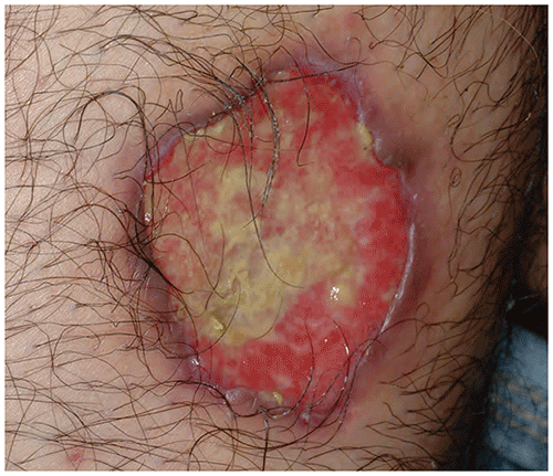
■Ulcers: deeper lesions extending into the dermis or below (Figure 1.11).
Figure 1.1. Café Au Lait Macules (Spots) in a Patient Who Has Neurofibromatosis Type 1.
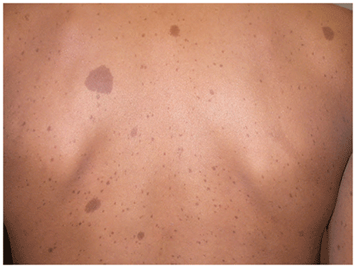
Figure 1.2. A Port-Wine Staina Vascular Patch.
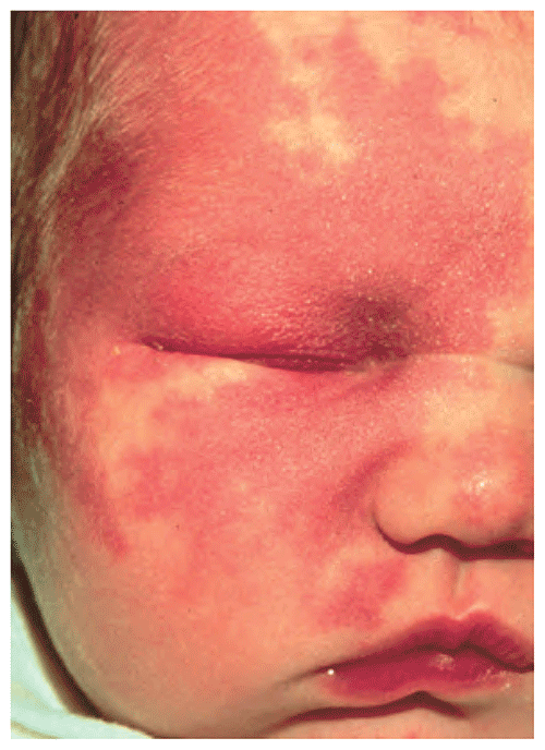
Figure 1.3. Molluscum Contagiosum. There are Erythematous and Skin-Colored Papules.
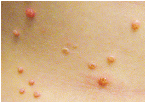
Figure 1.4. Nodules that are Neurofibromas in a Patient Who Has Neurofibromatosis Type 1.
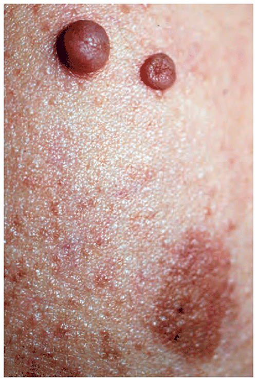
Figure 1.5. Pink Wheals in a Patient Who Has Urticaria.
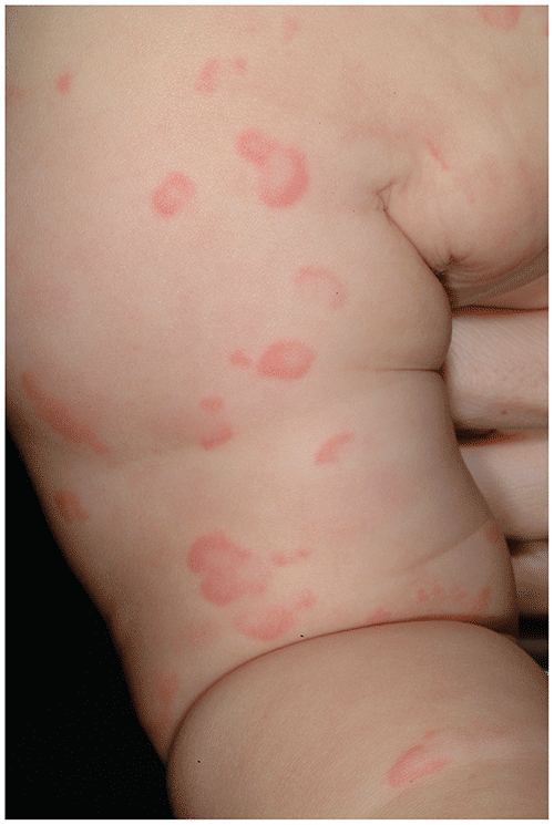
Figure 1.6. Scaling Plaques, Plateau-Like Lesions, are Observed in Psoriasis.
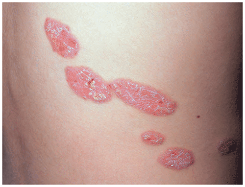
Figure 1.7. Vesicles, as Seen Here in Varicella, are Filled with Serous or Clear Fluid.
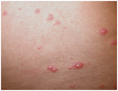
Figure 1.8. Bullae, Filled with Clear Fluid, are Observed in Chronic Bullous Disease of Childhood.
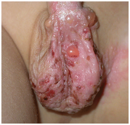
Figure 1.9. Pustules are Filled with Purulent Material. This Infant Has Congenital Cutaneous Candidiasis.
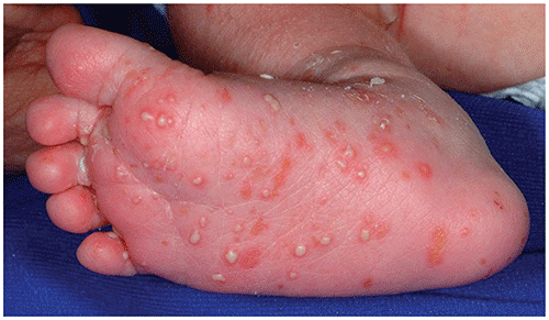
Figure 1.10. Erosions, as Seen in This Infant Who Has Acrodermatitis Enteropathica, Indicate a Superficial Loss of Epidermis.
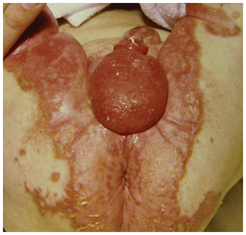
Figure 1.11. An Ulcer Occurs When There Has Been Loss of Epidermal and Dermal Tissues. In the Patient Shown Here, the Ulcer is the Result of Pyoderma Gangrenosum.
Distribution of Lesions
Certain disorders are characterized by unique patterns of lesion distribution. For example
▶Atopic dermatitis in children and adolescents typically involves the antecubital or popliteal fossae.
▶Seborrheic dermatitis in adolescents commonly involves not only the scalp but also the eyebrows and nasolabial folds.
▶Lesions of psoriasis are often seen in areas that are traumatized, such as the extensor surfaces of the elbows and knees.
▶Acne is limited to the face, back, shoulders, and chest, sites of the highest concentrations of pilosebaceous follicles.
Arrangement of Lesions
The arrangement of lesions also may provide a clue to diagnosis. Some examples include
▶Linear: allergic contact dermatitis due to plants (eg, poison ivy) (Figure 1.12), lichen striatus, and incontinentia pigmenti; may also occur in epidermal nevi, psoriasis, and warts.
▶Grouped: herpes simplex virus infection (Figure 1.13), warts, molluscum contagiosum, and microcystic lymphatic malformation.
▶Dermatomal: herpes zoster (Figure 1.14).
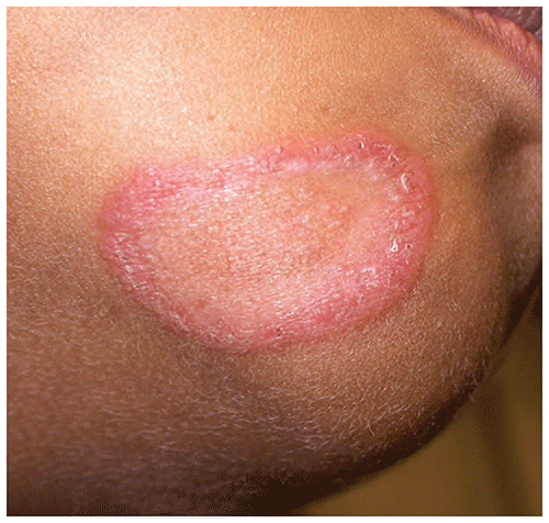
▶Annular (ie, ring-shaped with central clearing): tinea corporis (Figure 1.15), granuloma annulare, erythema migrans, and lupus erythematosus.
Figure 1.12. A Linear Arrangement of Papules or Vesicles Often Occurs in Allergic Contact Dermatitis Due to Poison Ivy.
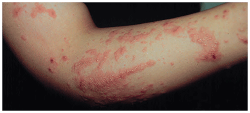
Figure 1.13. Grouped Vesicles are Characteristic of Herpes Simplex Virus Infection on the Skin.
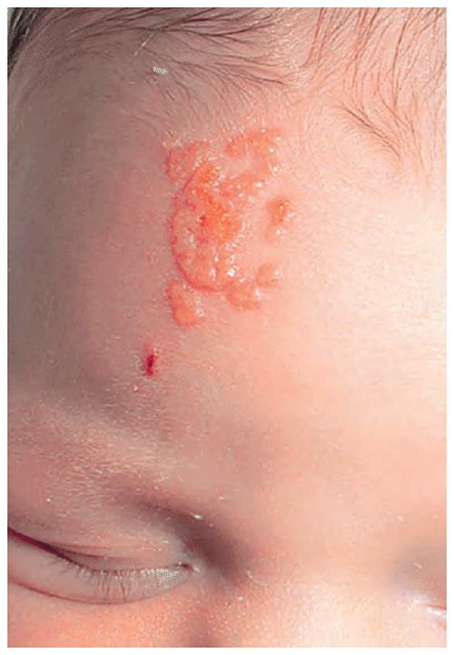
Figure 1.14. The Lesions of Herpes Zoster Appear in a Dermatomal Distribution.
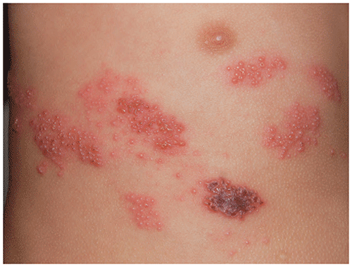
Figure 1.15. An Annular (Ring-Shaped with Central Clearing) Plaque is Typical of Tinea Corporis.
Color
▶Erythematous: pink or red. When erythematous lesions are observed, it is important to note whether they blanch. If the red cells are within vessels, as occurs in urticaria, compression of the skin forces the cells into deeper vessels, and blanching occurs. However, if the cells are outside vessels, as occurs in forms of vasculitis, blanching will not occur. Non-blanching lesions are termed petechiae, purpura, or ecchymoses. Also note that in individuals with skin of color, erythema may be more difficult to appreciate.
▶Hyperpigmented: tan, brown, or black.
▶Hypopigmented: amount of pigment decreased but not entirely absent (as seen with postinflammatory pigmentary alteration).
▶Depigmented: all pigment absent (as occurs in vitiligo).
Secondary Changes
Alterations in the skin that may accompany primary lesions include
▶Excoriation: a superficial loss of skin (ie, an erosion) caused by scratching, picking, or rubbing.
▶Crusting: dried fluid; commonly seen after rupture of vesicles or bullae (as occurs with the honey-colored crust of impetigo).
▶Scaling: epidermal fragments that are characteristic of several disorders, including fungal infections (eg, tinea corporis) and psoriasis.
▶Atrophy: an area of surface depression due to absence of the epidermis, dermis, or subcutaneous fat; atrophic skin often is thin and wrinkled. Examples include steroid atrophy, morphea, and atrophoderma.
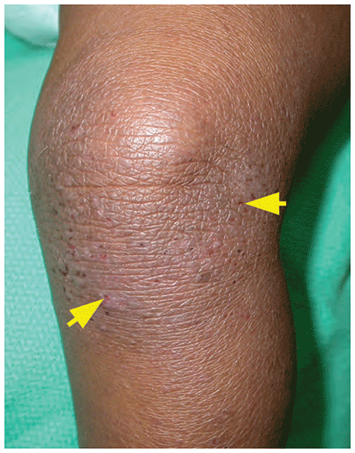
▶Lichenification: thickening of the skin from chronic rubbing or scratching (as occurs in atopic dermatitis); as a result, typical skin markings and creases appear more prominent (Figure 1.16).
Figure 1.16. Lichenification. The Typical Skin Markings are Very Prominent Due to Chronic Scratching. Also Note the Tiny Erosions (Arrows), Some of Which Have Formed Crusts.
Appearance Differences in Skin of Color
▶The degree of skin pigmentation often influences the appearance of certain dermatologic disorders. In patients with darker skin tones, erythema may be difficult to appreciate or virtually absent (Figure 1.17) or may appear violaceous, brown, gray, blue, or black.
▶Postinflammatory hypo- or hyperpigmentation occurs commonly in patients with darker skin tones (Figures 1.17 and 1.18). These pigmentary disturbances may be troubling for patients and may take months to resolve.
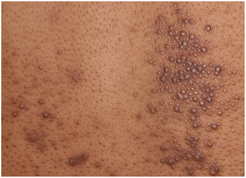
▶In patients with darker skin tones, lesion morphology also may differ from that seen in patients with less pigmentation. In atopic dermatitis and pityriasis rosea, for example, lesions may be papular (often follicular in nature, resembling goose bumps) rather than patches or plaques (Figures 1.19 and 1.20). In patients with atopic dermatitis, somewhat larger, flat-topped (lichenoid) papules may be observed (Figure 1.21). These papules may occasionally appear more violaceous.
▶Some additional disorders that may appear different in patients with darker skin tones compared with patients with lighter skin tones include
■Capillary malformations: port-wine stains may appear more brown or reddish-brown and may be mistaken for café au lait macules.
■Infantile hemangiomas: superficial infantile hemangiomas may appear violaceous rather than bright red, and deep infantile hemangiomas may lack the characteristic blue color seen in patients with lighter skin tones.
■Morbilliform drug eruptions: in patients with darker skin tones the erythematous macules and papules may be difficult to appreciate.
■Psoriasis: plaques may appear violaceous or hyperpigmented.
■Urticaria: wheals may appear more skin-colored or pale rather than erythematous.
■Viral exanthems: in patients with darker skin tones the erythematous macules may be difficult to appreciate.
Figure 1.17. Subtle Erythema and Postinflammatory Hypopigmentation in an Infant with Skin of Color Who Has Atopic Dermatitis.
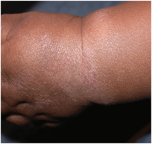
Figure 1.18. Postinflammatory Hyperpigmentation in an Adolescent with Skin of Color Who Has Acne.
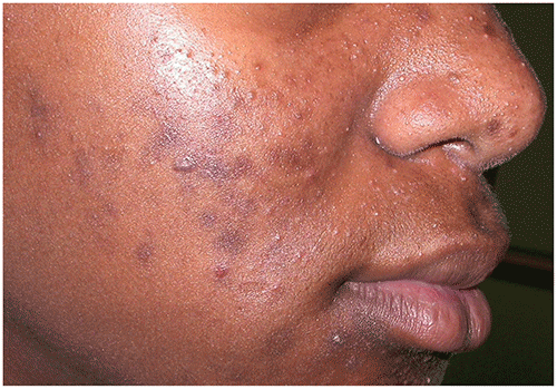
Figure 1.19. Tiny Follicular Papules in an Adolescent with Skin of Color Who Has Atopic Dermatitis. Reproduced with Permission from Krowchuk Dp. Practical Aspects of the Diagnosis and Management of Atopic Dermatitis. Pediatr Ann. 1987;16(1):57–66.
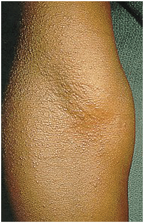
Figure 1.20. Abdomen of a Patient with Skin of Color Who Has Pityriasis Rosea. There are Tiny Follicular Papules and Oval Thin Plaques with Erythema that is More Subtle and Associated Central Hyperpigmentation in Several Lesions.
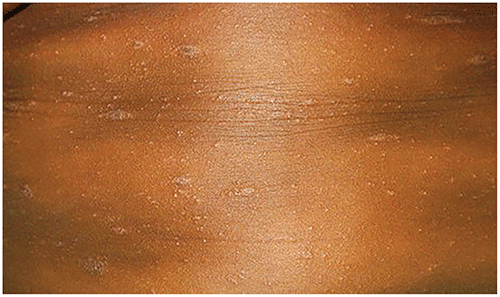
Figure 1.21. In Some Patients with Skin of Color Who Have Atopic Dermatitis, Areas of Disease Activity May Appear as Slightly Larger, Flat-Topped (Lichenoid), Violaceous Papules.