▶Acquired melanocytic nevi are common.
▶Begin to appear after 2 to 3 years of age.
▶Increase in number and reach a peak during the third decade.
▶Often disappear with advancing age.
▶Pigmented macules, papules, and plaques with variable surface changes.
▶Sometimes classified based on appearance.
■Junctional nevus: uniformly hyperpigmented (often brown) macule (Figure 69.1).
■Compound nevus: uniformly hyperpigmented (often brown) slightly elevated papule (Figure 69.2).
■Intradermal nevus: often light brown to flesh-colored and elevated (Figure 69.3).
▶Variants of acquired nevi
■Halo nevi
– Acquired nevi that develop a surrounding ring of hypopigmentation or depigmentation (Figure 69.4).
– Likely represents an immunologic response to melanocytes; often coexists with vitiligo (and may precede or follow this diagnosis).
– Nearly always benign in children; refer for evaluation if the ring of hypopigmentation is incomplete or the nevus is atypical using ABCDE criteria (see
When to Worry or Refer
section).– Nevus and hypopigmentation ultimately resolve.
■Atypical nevi
– Often larger (5–12 mm) than common acquired nevi; have irregular and ill-defined borders (Figure 69.5).
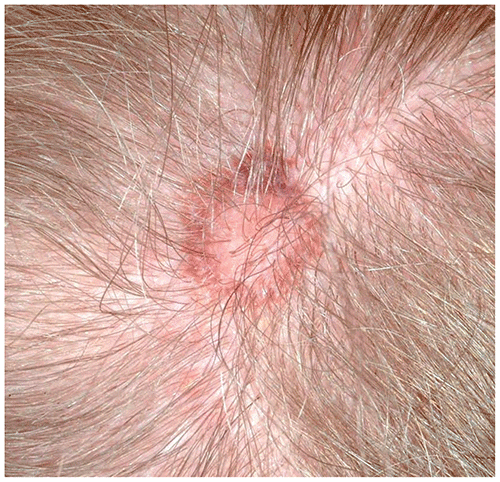
– Color often is variegated with shades of brown, tan, or pink (Figure 69.6).
– Individuals who have large numbers of atypical nevi or those who have a family history of melanoma in first-degree relatives have an increased risk of developing melanoma.
■“Eclipse” nevi
– Nevus with central elevation simulating the appearance of a sunny-side up fried egg (see Figure 69.6).
– Periphery often darker compared with the lighter central portions.
– This phenotype is common on the scalps of older children and teenagers, and these nevi tend to behave in benign fashion.
Figure 69.1. Junctional Nevus.
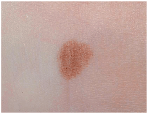
Figure 69.2. Compound Nevus.
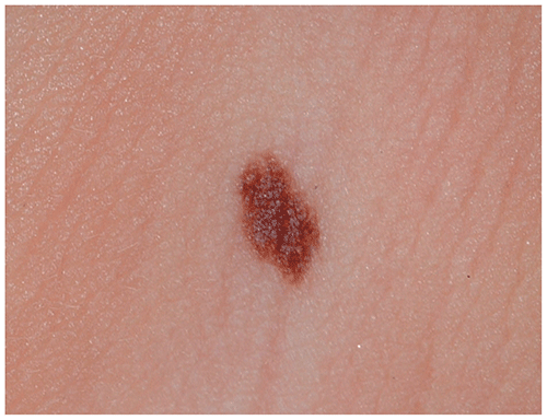
Figure 69.3. Intradermal Nevus.
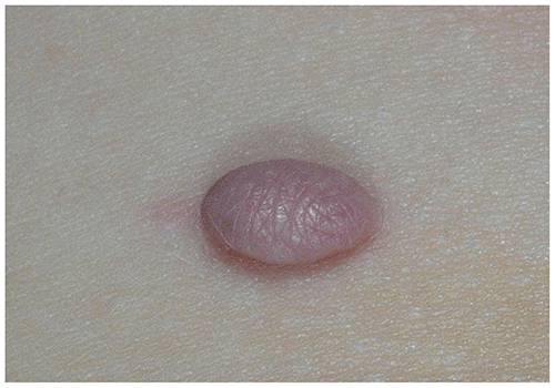
Figure 69.4. A Halo Nevus is an Acquired Nevus with a Ring of Surrounding Hypopigmentation or Depigmentation.
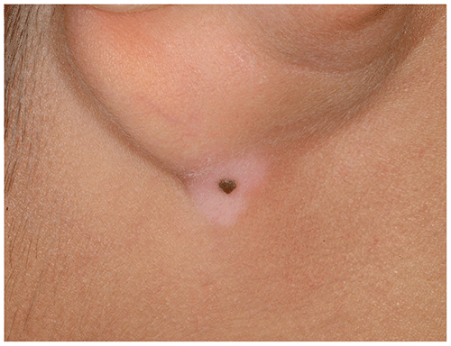
Figure 69.5. Atypical Nevi are Often Larger (Often 5–12 mm in Diameter) and Have Irregular Borders.
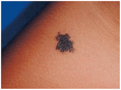
Figure 69.6. This Benign “eclipse” Nevus Reveals Pink and Brown Color; the Lighter Center is Elevated, Causing the Lesion to Have the Appearance of a Sunny-Side Up Fried Egg.
Look-alikes
| Disorder | Differentiating Features |
|---|
| Ephelides | Small, hyperpigmented macules located in sun-exposed areas such as the face, upper chest, shoulders, and back. Become darker following sun exposure. Often fade when sun exposure is minimal. Unlike melanocytic nevi, have no change in surface texture. No malignancy potential.
|
| Lentigines | Small, hyperpigmented macules not limited to sun-exposed areas. Unlike melanocytic nevi, have no change in surface texture. No malignancy potential.
|
| Café au lait macules | |
▶Benign-appearing nevi that are asymptomatic do not require removal.
▶Rapidly changing, symptomatic, or significantly atypical nevi must be assessed for possible malignant transformation.
▶Familial atypical mole/melanoma syndrome should be considered in a patient who has atypical (ie, dysplastic) moles and several family members with atypical (ie, dysplastic) nevi and at least one relative with melanoma. These patients require close surveillance to assess for the development of melanoma.
▶Ordinary acquired nevi are typically inconsequential (unless in a cosmetically compromising region); however, all nevi should be monitored using ABCDE criteria (see
When to Worry or Refer
section).▶Atypical nevi may imply an increased risk for the development of melanoma.
▶Melanoma is the malignant neoplasm of melanocytes that may arise de novo or from preexisting nevus. Consider the possibility of melanoma when a nevus exhibits any of the following ABCDE criteria:
■Asymmetry
■Border irregularity
■Color variation (especially red, blue, black)
■Diameter larger than about 6 mm
■Evolving lesion that is changing quickly
▶Recently, it has been suggested that applying these traditional ABCDE criteria in addition to modified ABCD criteria may facilitate earlier detection of melanoma in children. The modified ABCD criteria (and explanation) are as follows:
■Amelanotic (as pediatric melanoma may be nonpigmented).
■Bleeding, bump (bleeding lesions; lumps or bumps [ie, papulonodules or papules]).
■Color uniformity (as the color may be consistent and uniform throughout the lesion).
■De novo, any diameter (new papular lesions; lesions may be <6 mm).
▶Refer patients who have atypical nevi and a family history of atypical nevi or melanoma to a dermatologist.
▶Refer patients who have atypical-appearing halo nevi (eg, those with an incomplete ring of hypopigmentation or depigmentation or an atypical appearance according to ABCDE criteria) to a dermatologist.