▶Common dermatophyte infection of the scalp; in the United States, Trichophyton tonsurans, Microsporum canis, and Microsporum audouinii are responsible for most cases.
▶T tonsurans is responsible for more than 90% of US infections.
▶For reasons unknown, Black children are disproportionately affected.
Three patterns of infection may be observed.
▶Alopecia
■One or more round or oval patches of partial to complete alopecia with associated scaling (Figure 37.1).
■Infections caused by T tonsurans cause hairs to break at the scalp, resulting in black dot hairs (the remnants of hairs remaining within the follicle) (see Figure 37.1).
■Infections caused by Microsporum species cause hairs to break further from the scalp, resulting in incomplete alopecia; black dot hairs are absent.
▶Seborrheic
■Mimics seborrheic dermatitis (ie, dandruff) with patchy or diffuse whitish to gray scale (Figure 37.2).
■Alopecia may be subtle.
▶Inflammatory: When an inflammatory response to the infecting agent occurs, patients may develop
■Papules, pustules, and crusting that may mimic bacterial folliculitis.
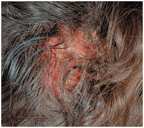
■A tender, boggy mass known as a kerion (Figure 37.3).
▶All forms of tinea capitis, but particularly inflammatory forms, may produce suboccipital or posterior cervical lymphadenopathy.
Figure 37.1. Tinea Capitis. A Well-Defined Patch of Alopecia within Which are Scale, Black Dot Hairs (Yellow Arrows), and Pustules (Red Arrow).
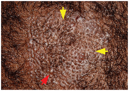
Figure 37.2. Diffuse Scaling of the Scalp is Observed in the Seborrheic Form of Tinea Capitis.
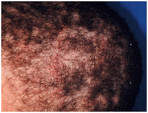
Figure 37.3. A Kerion is a Tender, Boggy Mass Located on the Scalp.
Look-alikes
In each of the conditions listed herein, a potassium hydroxide preparation or fungal culture would fail to confirm the presence of fungal infection.
| Disorder | Differentiating Features |
|---|
| Alopecia areata | Round or oval patches of alopecia that lack scaling, inflammation, or black dot hairs. Nail pitting often present.
|
| Trichotillomania | Often ill-defined patches of alopecia within which hairs are of differing lengths. Petechiae or hemorrhagic crusts may be present if hairs pulled from the scalp. Scaling and black dot hairs absent. History of hair manipulation may be offered by family (but not always).
|
| Bacterial folliculitis | Alopecia and scaling absent. Culture positive for Staphylococcus aureus. Note: In patients who have tinea capitis, S aureus often can be cultured from the scalp (although the pustules themselves may be sterile).
|
| Bacterial abscess | Less likely to produce alopecia than a kerion. Scaling absent. Culture of contents usually reveals S aureus or other bacterial organisms. Note: In patients who have tinea capitis, S aureus often can be cultured from the scalp (although the pustules themselves may be sterile).
|
| Traction alopecia | Traction on hair may produce alopecia localized to areas where hair is parted. Folliculitis may occur, but scaling and black dot hairs absent. History of tight braids or ponytails often present, with hair thinning in peripheral zones.
|
| Seborrheic dermatitis | |
▶The diagnosis usually is made clinically and supported by laboratory testing.
■The presence of occipital lymphadenopathy and alopecia, or lymphadenopathy and scaling, are highly predictive of tinea capitis.
▶A potassium hydroxide preparation performed on infected hairs will reveal spores within the hair shaft (ie, endothrix infection as caused by T tonsurans) (Figure 37.4) or on the surface of hairs (ie, ectothrix infection as caused by Microsporum species).
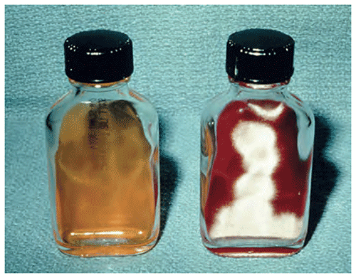
▶Culture (the criterion standard for diagnosis) of scale or hair fragments on dermatophyte test medium (or other suitable medium) confirms the diagnosis (Figure 37.5). Consider performing a culture when diagnostic uncertainty exists; some also use culture to confirm a mycological cure prior to discontinuation of therapy.
■Specimens for culture may be obtained with a Cytobrush, toothbrush, or premoistened cotton-tipped applicator.
■Sensitivity of culture is high, even with delay in inoculation of medium due to transportation of specimen to laboratory.
▶Wood lamp examination is useful only in ectothrix infections (ie, those caused by Microsporum species). In such cases, infected hairs will fluoresce. Infections caused by T tonsurans (>90% of infections) do not fluoresce.
Figure 37.4. Tinea Capitis Caused by Trichophyton Tonsurans Produces an Endothrix Infection. The Infected Black Dot Hair is Filled with Arthrospores, the Spherical Objects Shown Here.
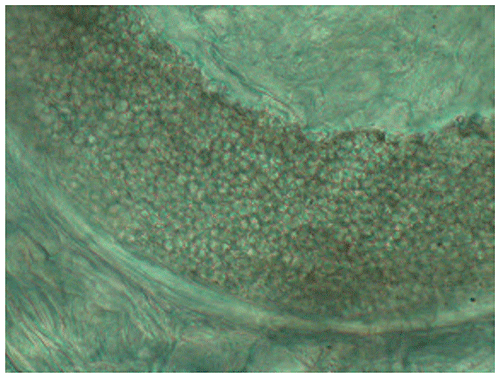
Figure 37.5. The Diagnosis of Tinea Capitis May be Confirmed by Performing a Fungal Culture. Uninoculated Medium is Yellow (Left). Within 2 Weeks of Inoculation with Scale or Black Dot Hairs Scraped from the Scalp, There is Fungal Growth and the Medium Turns Red (Right).
▶Oral therapy is required. A summary of treatment options is provided in Table 37.1.
■For many years, griseofulvin was considered the drug of choice for the treatment of tinea capitis. However, when compared with newer agents discussed as follows, it requires longer therapy and offers lower efficacy. Griseofulvin is administered at a dose of 20 to 25 mg/kg/d of the micro-sized preparation or 15 mg/kg/d of the ultramicro-sized preparation. Patients should be treated for 6 to 8 weeks minimum. Laboratory monitoring is not necessary.
■Terbinafine, fluconazole, and itraconazole have proven effective in treating tinea capitis (terbinafine is US Food and Drug Administration approved for tinea capitis in patients 4 years or older, while both fluconazole and itraconazole are not approved for this indication). Laboratory monitoring is considered with the use of these antifungal therapies.
– These agents (particularly terbinafine) often are used to treat patients in whom griseofulvin fails. Many practitioners consider terbinafine as first-line therapy.
– Terbinafine is less effective than griseofulvin in the treatment of tinea capitis caused by Microsporum species.
– Fluconazole is the only systemic antifungal agent approved for use in patients younger than 2 years, although not specifically for tinea capitis.
▶The use of an adjunctive antifungal shampoo containing selenium sulfide (1% or 2.5%) or ketoconazole 2% twice weekly will kill surface spores and, possibly, reduce spread of infection to others. The agent should be used for at least 2 weeks.
▶Some authors recommend the addition of oral prednisone (eg, for 1–3 weeks) to the treatment regimen in patients who have severe inflammatory tinea capitis (ie, a kerion).
▶Incision and drainage of a kerion is not indicated.
▶Patients should be seen in follow-up 1 to 2 months after beginning therapy to assess response.
▶Children should not be excluded from school once therapy is begun. Some experts recommend that asymptomatic family members use an antifungal shampoo, although evidence is lacking regarding the efficacy of this strategy. If a dog or cat is suspected to be the source of infection, the animal should be evaluated and treated if appropriate.
Table 37.1. Recommended Therapy for Tinea Capitis
| Drug | Dosage | Duration |
|---|
| Griseofulvin microsize (liquid 125 mg/5 mL) | 15–25 mg/kg/d (max 1 g/d) | 6–8 wk; continue until clinically clear
FDA approved for children ≥2 y |
| Griseofulvin ultramicrosize (tablets of varying size) | 10–15 mg/kg/d (max 750 mg/d) | 6–8 wk; continue until clinically clear
FDA approved for children ≥2 y |
| Terbinafine tablets (250 mg) a | 4–6 mg/kg/d (max 250 mg); or
10–20 kg: 62.5 mg
20–40 kg: 125 mg
>40 kg: 250 mg | Trichophyton tonsurans: 4–6 wk
Microsporum canis: 8–12 wk
Not FDA approved for the treatment of tinea capitis in children |
| Terbinafine granules (125 mg and 187.5 mg) b | <25 kg: 125 mg
25–35 kg: 187.5 mg
>35 kg: 250 mg | Trichophyton tonsurans: 4–6 wk
Microsporum canis: 8–12 wk
FDA approved for children ≥4 y |
| Fluconazole c (liquid 10 mg/mL, 40 mg/mL; tablet 50 mg, 100 mg, 150 mg, and 200 mg) | 6 mg/kg/d (max 400 mg/d) | 3–6 wk depending on severity
Not FDA approved for the treatment of tinea capitis in children |
| Itraconazole solution c (10 mg/mL) | 3 mg/kg/d (max 600 mg/d) | 2–4 wk or longer, depending on severity
Not FDA approved for the treatment of tinea capitis in children |
| Itraconazole capsule (65 mg, 100 mg) | 5 mg/kg/d (max 600 mg/d) | 2–4 wk or longer, depending on severity
Not FDA approved for the treatment of tinea capitis in children |
Abbreviations: FDA, US Food and Drug Administration; max, maximum.
Some experts use “higher” dosing listed for terbinafine granules instead.
Terbinafine granules have been discontinued in the in the United States.
See Red Book: 2024–2027 Report of the Committee on Infectious Diseases Antifungal Drugs for Systemic Fungal Infections and Recommended Doses of Parenteral and Oral Antifungal Drugs, for adverse reactions and therapeutic drug monitoring recommendations.
From American Academy of Pediatrics. Tinea capitis. In: Kimberlin DW, Banerjee R, Barnett ED, et al, eds. Red Book: 2024–2027 Report of the Committee on Infectious Diseases. 33rd. American Academy of Pediatrics; 2024:854–858.
▶Although Staphylococcus aureus may be cultured from the scalp of children who have tinea capitis, antibiotic treatment usually is unnecessary.
▶If clinical evidence of secondary bacterial infection is present, an antistaphylococcal antibiotic should be prescribed.
▶The prognosis is excellent. With treatment, alopecia resolves in nearly all patients (those who have a large kerion occasionally will experience permanent alopecia).
▶Reinfection is common in children who share potential fomites (eg, hats, scarves, headgear, earphones, combs, brushes) or those who are reexposed to infection (from children or pets).