section name header

Basics

Outline


BASICS

Definition!!navigator!!

Localized ulcerative inflammation of the right dorsal colon often associated with administration of NSAIDs, particularly phenylbutazone or flunixin meglumine; dehydration increases the risk of drug toxicity.

Pathophysiology!!navigator!!

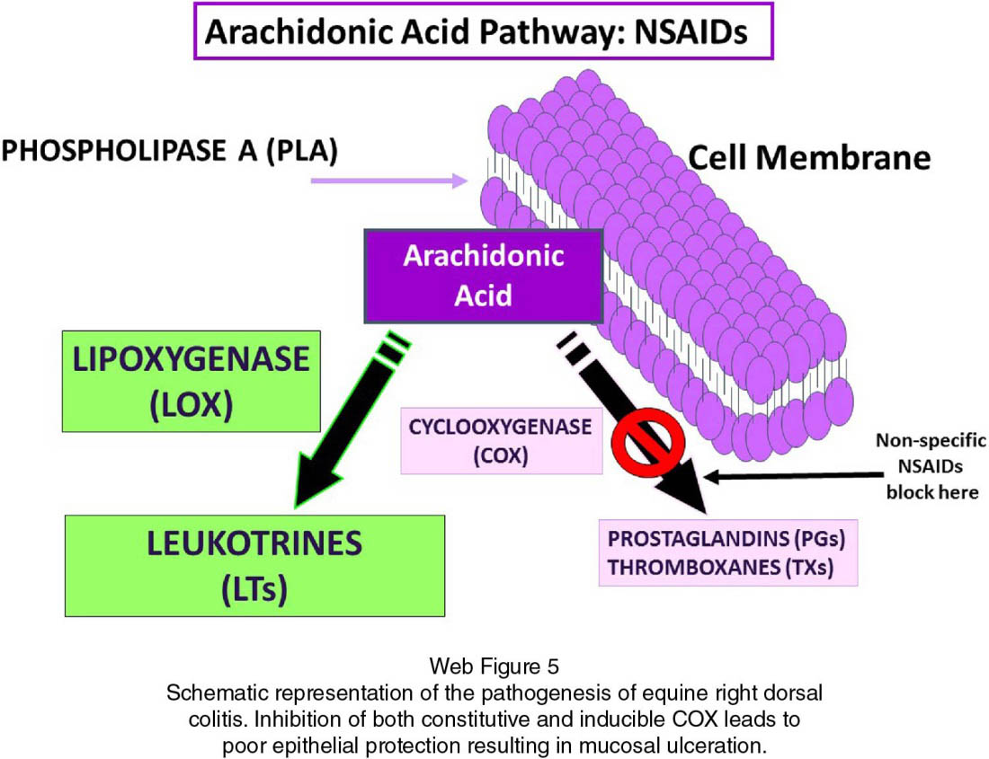
  • Nonspecific NSAIDs including phenylbutazone and flunixin meglumine inhibit COX activity (Web Figure 1)

    as a competitive antagonist for both constitutive COX-1, responsible for the production of PGs involved in physiologic homeostatic functions, and for inducible COX-2, upregulated with inflammation. Intestinal mucosal cell production of PGE2 and PGF2α is decreased by the inhibition of COX-1. This results in the loss of the PG-mediated protective effects on the intestinal mucosa. When the intestinal mucosa integrity is sufficiently compromised, local bacterial invasion of the mucosa, luminal endotoxin absorption, and plasma protein leakage into the intestinal lumen may occur
  • NSAID toxicity is potentiated in dehydrated or hypovolemic horses because the normal protective vascular changes guarding from reduced blood flow are inhibited
  • The development of RDC in absence of previous NSAID administration suggests that the condition may be multifactorial

Systems Affected!!navigator!!

Gastrointestinal Tract

  • The right dorsal colon is primarily affected. Oral and gastric ulcerations may also develop from NSAID toxicity
  • Histologically, lesions are characterized by multifocal to coalescing ulcerations in the wall of the right dorsal colon (Web Figure 2)
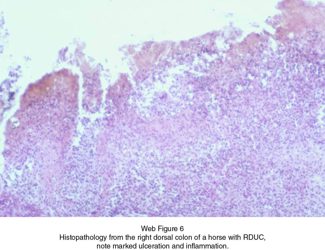
  • Subacute lesions are characterized by a fibrinonecrotic ulcerative colitis
  • In chronic cases, fibrous connective tissue is present in the lamina propria underlying the ulcerated mucosa. Colonic stenosis with ingesta impaction and subsequent necrosis and rupture of the colon can occur

Genetics!!navigator!!

N/A

Incidence/Prevalence!!navigator!!

RDC, now preferably called RDUC (U ulcerative), may be associated with the administration of phenylbutazone or other NSAID therapy at the manufacturer's recommended daily dose. The variable toxicity has been attributed to individual variation in response to NSAIDs, duration of treatment, diet composition, health status, age, and hydration status.

Geographic Distribution!!navigator!!

N/A

Signalment!!navigator!!

Although there is no evidence to demonstrate overrepresentation for any particular age or breed, most reports involve miniature horses, ponies, and young performance horses.

Signs!!navigator!!

General Comments

Most cases have a history of administration of oral or parenteral NSAIDs with phenylbutazone being the most commonly used. RDUC can also result following high-dose administration of flunixin meglumine or in combination with phenylbutazone. NSAID therapy is most commonly implemented for treatment of chronic or severe musculoskeletal conditions (i.e. laminitis).

Historical Findings

  • NSAID toxicity cases commonly present with concurrent systemic dehydration, which may result from either systemic illness or an inability to maintain proper hydration
  • Horses with acute disease commonly have clinical signs that include colic, depression, lethargy, partial or complete anorexia, fever, and diarrhea
  • Horses with chronic disease may demonstrate intermittent colic, soft feces, weight loss, and ventral edema

Physical Examination Findings

  • Acute disease is commonly associated with signs of colitis including watery diarrhea, severe dehydration, severe systemic illness, injected mucous membranes and/or marked endotoxemia
  • Horses with chronic disease may have formed or soft “cow pie” feces, edema, and abnormal vital parameters that include often elevated heart rate

Causes!!navigator!!

NSAID toxicity results from high-dose therapy, with individual horses having the ability to tolerate higher NSAID administration doses. It is not clear why ulcerative lesions are localized in the right dorsal colon.

Risk Factors!!navigator!!

Dehydration in combination with NSAID administration are predisposing factors for RDUC.

Diagnosis

Outline


DIAGNOSIS

Differential Diagnosis!!navigator!!

  • Other causes of infectious colitis including salmonellosis, Clostridium difficile infection, and Potomac horse fever should be considered
  • Chronic NSAID toxicity may have similarities to other forms of PLE including cyathostomiasis, chronic inflammatory (infiltrative) bowel disease, alimentary lymphoma, Lawsonia intracellularis, chronic salmonellosis, gastric ulcers, or sand enteropathy. Definitive diagnosis is based on the history of NSAID administration, physical examination findings, and hematologic/biochemical evidence of marked PLE

CBC/Biochemistry/Urinalysis!!navigator!!

CBC

With acute disease hematologic abnormalities often include neutropenia with mild to moderate toxic neutrophils in combination with a regenerative or a degenerative left shift. An increased packed cell volume is frequently observed. Patients that present with more chronic disease may have a less pronounced neutropenia, but neutrophil morphology may still reveal toxicity.

Biochemistry Profile

Serum biochemical abnormalities often include marked hypoproteinemia, hypochloremia, azotemia and metabolic acidosis. Hypoproteinemia may be observed despite hemoconcentration and will worsen with rehydration. Hypoalbuminemia is common, but when total protein concentration is <4.5 g/dL (45 g/L), panhypoproteinemia is typically observed.

Other Laboratory Tests!!navigator!!

Abdominal Paracentesis

Elevation of total solids concentration and nucleated cell count may be observed.

Fecal Occult Blood Test

Generally positive—this should be performed before rectal examination to avoid any complications with interpretation of test result.

Imaging!!navigator!!

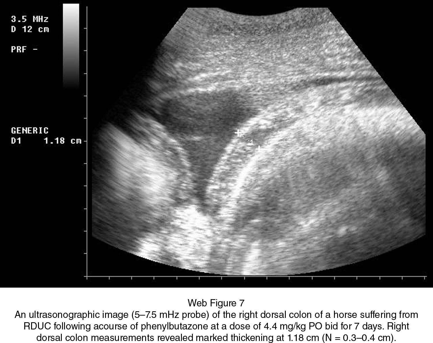
  • Transabdominal ultrasonography at the right 10th to 13th intercostal spaces can be used in standing horses to image the right dorsal colon. Horses with RDUC have significantly greater colonic wall thickness than healthy horses (N = 0.3–0.4 cm). The right dorsal colon of affected horses also has a prominent hypoechoic layer associated with submucosal edema and inflammatory infiltrates (Web Figure 3)

  • Gastroscopy—horses with RDUC commonly have concurrent gastric and potentially duodenal ulcers. Diagnosis of gastric ulceration will impact treatment protocols

Other Diagnostic Procedures!!navigator!!

Exploratory Celiotomy/Laparoscopy

Rarely indicated. Gross examination reveals marked edema, thickening, and/or when the reduced luminal diameter of the intestinal tract is restricted to right dorsal colon.

Intestinal Biopsy

Definitive diagnosis of the condition is made by histopathologic examination of a biopsy of the right dorsal colon obtained during celiotomy or at necropsy.

Treatment

Outline


TREATMENT

Aims!!navigator!!

The treatment consists of discontinuation of NSAID therapy in combination with appropriate supportive therapy.

Appropriate Health Care!!navigator!!

The majority of patients with RDUC require hospitalization to receive appropriate medical supportive therapy.

Nursing Care!!navigator!!

Acute RDC

Horses with acute disease are treated with supportive treatment, including IV fluids (LRS), systemic broad-spectrum antibiotics, and analgesics. All NSAIDs should be discontinued. When hypoproteinemia is severe (albumin <1.5 g/dL; 15 g/L), plasma transfusion should be administered.

Chronic RDC

  • Horses with chronic disease are managed with low-residue feeding and discontinuation of all NSAIDs
  • Dietary management consists of frequent feeding (4–6 meals) of complete, pelleted (low residue) diet that contains 30% dietary fiber and 14% protein. Pelleted feed is advocated because it decreases the mechanical and physiologic load on the large colon. Long-stem roughage is eliminated or restricted to small amounts of fresh grass for at least 3 months. Low-residue diet is fed according to the manufacturer's recommendations. The diet change is gradually completed over a period of approximately a week.

Activity!!navigator!!

Managements to decrease stress include discontinuing or decreasing or eliminating work such as strenuous exercise.

Surgical Considerations!!navigator!!

Surgical intervention indicated when the RDUC cannot be controlled with medical treatment evidenced by recurrent colic episodes. Surgery requires either bypassing or resecting the diseased RDC. Side-to-side colo-colostomy, between the proximal intact part of the RDC and the small colon, can be performed to bypass the diseased part of the RDC. End-to-end colo-colostomy after resection of the diseased RDC can also be performed. The prognosis for horses that undergo surgery is guarded.

Medications

Outline


MEDICATIONS

Drug(s) of Choice!!navigator!!

Supportive treatment for horses with acute disease includes:

  • LRS (60–120 mL/kg/24 h) for volume replacement
  • Broad-spectrum systemic antibiotics (potassium penicillin G 22 000 IU/kg IV QID and gentamicin sulfate 6.6 mg/kg IV daily or ceftiofur 2.2 mg/kg IV BID)
  • Analgesics such as butorphanol (0.02 mg/kg IV or as a constant rate infusion at 0.013 mg/kg/h IV in fluids) can be administered

Precautions!!navigator!!

  • NSAIDs such as phenylbutazone or flunixin meglumine should be avoided because they are involved in the pathogenesis of the disease
  • Aminoglycosides should be used with caution when clinical signs of severe dehydration (prerenal azotemia) are present owing to its potential nephrotoxicity

Follow-up

Outline


FOLLOW-UP

Patient Monitoring!!navigator!!

  • Continual hematologic monitoring until resolution of hypoproteinemia
  • Include overall vital parameters and during rehydration as well hematocrit and plasma protein concentrations. If plasma protein concentrations decrease below 4 g/dL (40 g/L) or albumin drops below 1.5 g/dL (15 g/L) plasma transfusion is indicated
  • Continued monitoring of total protein concentrations will provide evidence of colonic healing. Once the total protein, specifically albumin concentration, is near or within normal ranges dietary restrictions can be gradually eliminated
  • Colonic restitution and resolution of hypoproteinemia/hypoalbuminemia requires generally 3–6 months

Possible Complications!!navigator!!

  • Horses with acute disease can develop a severe diarrhea, endotoxemia with marked edema, laminitis, or renal disease
  • Chronic disease may be associated with progressive weight loss, hypoproteinemia, colic, colonic stricture, and persistently loose feces

Miscellaneous

MISCELLANEOUS

Abbreviations

  • COX = cyclooxygenase
  • LRS = lactated Ringer's solution
  • NSAID = nonsteroidal anti-inflammatory drug
  • PG = prostaglandin
  • PLE = protein-losing enteropathy
  • RDC = right dorsal colitis
  • RDUC = right dorsal ulcerative colitis

Suggested Reading

Cook VL, Blikslager AT. The use of nonsteroidal anti-inflammatory drugs in critically ill horses. J Vet Emerg Crit Care 2015;25:7688.

Jones SL, Davis J, Rowlingson K. Ultrasonographic findings in horses with right dorsal colitis: five cases (2000–2001). J Am Vet Med Assoc 2003;222:12481251.

Karcher LF, Dill SG, Anderson WI, King JM. Right dorsal colitis. J Vet Intern Med 1990;4:247253.

McConnico RS, Morgan TW, Williams CC, et al. Pathophysiologic effects of phenylbutazone on the right dorsal colon in horses. Am J Vet Res 2008;69:14961505.

Moses VS, Bertone AL. Nonsteroidal anti-inflammatory drugs. Vet Clin North Am Equine Pract 2002;18:2137.

Author(s)

Author: Elizabeth Davis

Consulting Editors: Henry Stämpfli and Olimpo Oliver-Espinosa

Acknowledgment: The author and editors acknowledge the prior contribution of Ludovic Bouré.