Author(s): Anne Elizabeth Johnson, MSN, RN, CWON
The words used to describe a wound must communicate the same thing to members of the health care team, insurance companies, regulators, the patient's family, and, ultimately, the patient himself. The best way to classify wounds is to use the basic system described here, which focuses on three categories of fundamental characteristics:
- Wound age
- Wound depth
- Wound color
Wound age
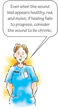
The first step in classifying a wound is to determine whether the wound is acute or chronic. Be careful; you can't base your determination solely on time because no set time frame specifies when an acute wound becomes chronic.
Acute
- New or relatively new wound
- Occurs suddenly
- Healing progresses in a timely, orderly, and predictable manner
- Typically heals by primary intention
- Examples: Surgical and traumatic wounds
Chronic
- May develop over time
- Healing has slowed or stopped
- Typically heals by secondary intention
- Examples: Pressure, vascular, and diabetic ulcers
Wound depth
Wound depth can be classified as partial thickness or full thickness.
In the case of pressure injuries/ulcers, wound depth allows you to stage the injury/ulcer according to the classification system developed by the National Pressure Ulcer Advisory Panel. (See Chapter 6, Pressure ulcers.)
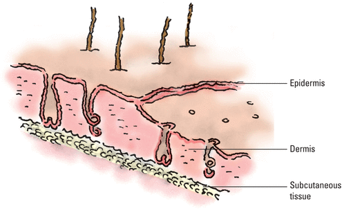
Partial-thickness wounds involve only the epidermis or extend into the dermis but not through it.
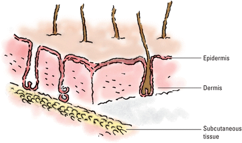
Full-thickness wounds extend through the dermis into tissues beneath and may expose adipose tissue, muscle, or bone.
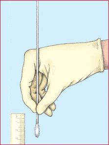
To measure the depth of a wound, you'll need gloves, a cotton-tipped swab, and a disposable measuring device. This method can also be used to measure wound tunneling or undermining.
- Put on gloves and gently insert the swab into the deepest portion of the wound.
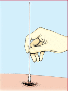
- Grasp the swab with your fingers at the point that corresponds to the wound's margin. You can carefully mark the swab where it meets the edge of the skin.
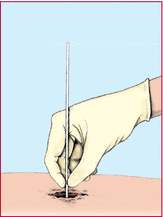
- Remove the swab and measure the distance from your fingers or from the mark on the swab to the end of the swab to determine the depth.
Wound color
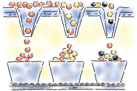
The Red-Yellow-Black Classification System is a commonly used approach that can help you determine how well a wound is healing and develop effective wound care management plans.
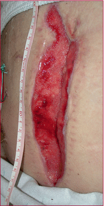
Red indicates normal healing. When a wound begins to heal, a layer of granulation tissue covers the wound bed. Granulation tissue is shiny and bright with a bumpy surface.

Granulation tissue in an abdominal wound
Fibrin leftover from the healing process usually appears as avascular yellow slough or dead tissue on the wound base. This slough, or soft necrotic tissue, provides a medium for bacterial growth.

Sacral pressure injury/ulcer with 75% of the surface area covered in yellow necrotic slough
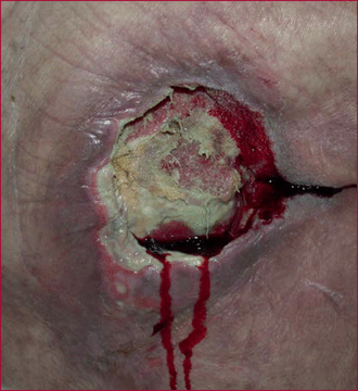
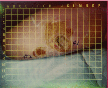
Diabetic foot ulcer with a slough-covered base and calloused edges.
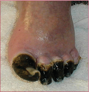
Black, the least healthy wound color, signals necrosis. Avascular dead tissue (known as eschar) slows healing and provides a site for microorganisms to proliferate.

Black ischemic toe ulcers
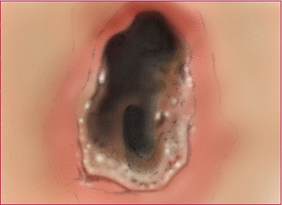
Black pressure ulcer
Best dressed Tailoring wound care to wound color
|
If you note two or even all three colors in a wound, classify the wound according to the least healthy color present. For example, if your patient's wound appears both red and yellow, classify it as a yellow wound.
