Author(s): Teresa J. Kelechi, PhD, RN, GCNS, CWCN, FAAN
The body's vascular system consists of:
- veins (carry blood toward the heart)
- arteries (carry blood away from the heart)
- lymphatic system (a separate circulatory system that collects waste products and delivers them to the venous system).
Veins
Veins carry deoxygenated blood back to the heart for reoxygenation.
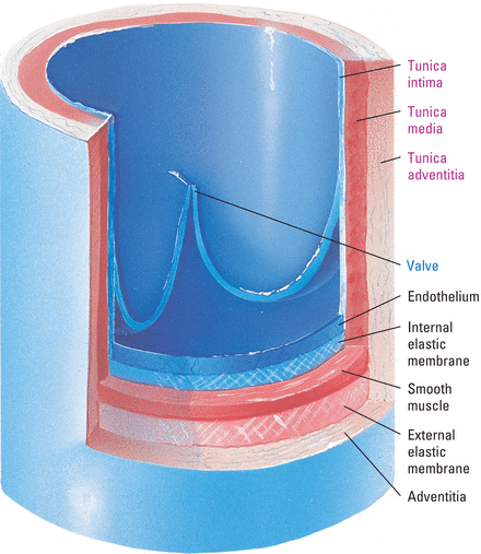
Vein walls have three layers. Compared to arteries of the same size, veins have thinner walls and wider diameters.
Veins have a unique system of cup-shaped valves that open toward the heart. The valves function to keep blood flowing in one direction—toward the heart.
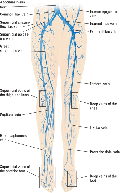
The illustration below shows the major veins in this part of the body.
The lower portion of the body contains three major types of veins and smaller vessels known as venules. Venules are very small veins whose purpose is to collect blood from the small capillaries in the arterial system.


Arteries
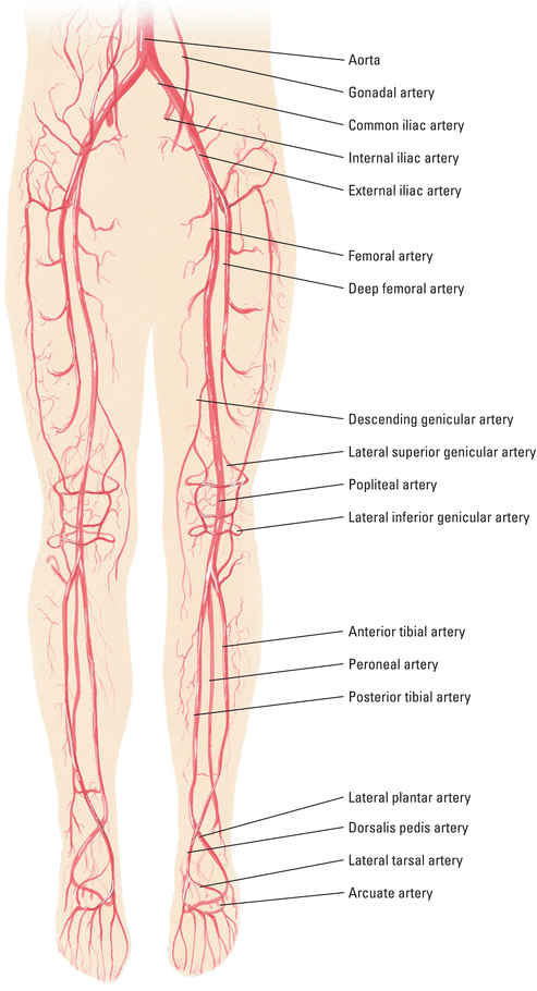
Arteries carry blood from the heart to every functioning cell in the body. The lower portion of the body receives its arterial flow through the abdominal aorta and the major arteries branching from it.
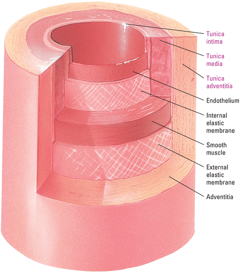
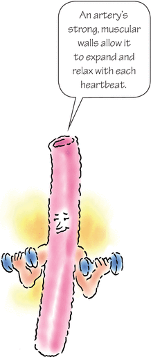
Like vein walls, artery walls have three layers.
This illustration identifies the major arteries in the lower portion of the body. Capillaries are the smallest of the blood vessels and make up the body's microcirculation. They connect arterioles and venules and facilitate movement of water, oxygen, carbon dioxide, and other substances between the tissues and blood around them.
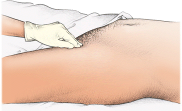
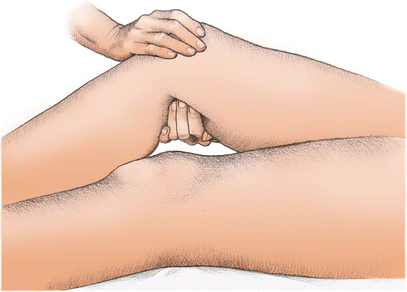
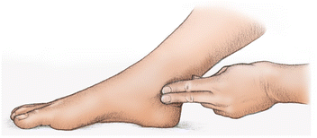
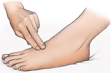
Assessing pulses is an effective way to evaluate arterial blood flow to the lower extremities. These illustrations show where to position your fingers when palpating for pulses of the lower extremities. Use your index and middle fingers to apply pressure.
Press firmly at a point inferior to the inguinal ligament. For obese patients, palpate in the groin crease, halfway between the pubic bone and hip bone.
Press firmly in the popliteal fossa at the back of the knee.
Apply pressure behind and slightly below the medial malleolus.
Place your fingers on the medial dorsum of the foot while the patient points his toes down. The pulse is difficult to palpate here and may seem absent in healthy patients. Sometimes, it helps to dorsiflex the foot to “pop up” the artery that lies between the first and second metatarsal bones.
In Doppler ultrasonography, high-frequency sound waves are used to assess blood flow. A handheld transducer, or probe, directs the sound waves into a vessel, where they strike moving red blood cells (RBCs). The frequency of the sound waves changes in proportion to the velocity of the RBCs. Doppler ultrasonography can be used to assess both arterial and venous blood flow.
Assessing arterial blood flow
- Apply a small amount of transmission gel to the ultrasound probe.
- Position the probe on the skin directly over the selected artery.
- Turn the instrument on and set the volume to the lowest setting.
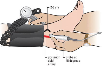
- To obtain the best signal, tilt the probe at a 45-degree angle from the artery, making sure that the gel is between the skin and the probe.
- Slowly move the probe in a circular motion to locate the center of the artery. Avoid pressing the probe too heavily on the artery to avoid compressing it.
- Listen for a triphasic, biphasic, or monophasic sound, which occurs when the Doppler signal isolates an artery.
- Count the signal for 60 seconds to determine the pulse rate.
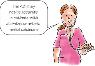
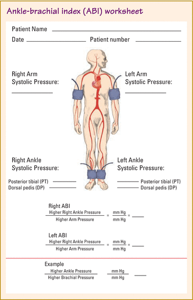
The ankle-brachial index (ABI) is a value derived from blood pressure measurements, which shows the progress or improvement of arterial disease. Each value in the index is a ratio of blood pressure measurement in the affected limb to the highest systolic pressure in the brachial arteries.
Steps
- Place the patient in a supine position with the legs at heart level.
- Measure and record both brachial blood pressures.
- Wrap the blood pressure cuff around one ankle, just above the malleolus, with the cuff bladder centered over the posterior tibial artery.
- Apply ultrasound transmission gel to a Doppler transducer.
- Hold the Doppler transducer over the dorsalis pedis artery at a 45-degree angle.
- Inflate the blood pressure cuff until the Doppler signal disappears.
- Slowly deflate the cuff until the Doppler signal returns. Record this pressure as the dorsalis pedis pressure.
- Repeat this same procedure over the posterior tibial artery. Record this pressure as the posterior tibial pressure.
- Calculate the ABI by dividing the highest ankle pressure by the highest brachial systolic pressure.
- Repeat the process on the contralateral limb.
Interpretation of results
- ABI > 0.9 = Normal
- ABI 0.71 to 0.9 = Mild arterial insufficiency
- ABI 0.41 to 0.7 = Moderate arterial insufficiency
- ABI 0 to 0.40 = Severe arterial insufficiency
Lymphatic system
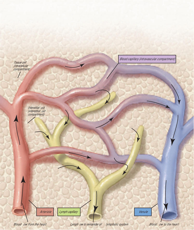
The lymphatic system is a vascular network that drains lymph (a protein-rich fluid similar to plasma) from body tissues and intravascular compartments and returns it to the venous system.
Lymphatic system and drainage route
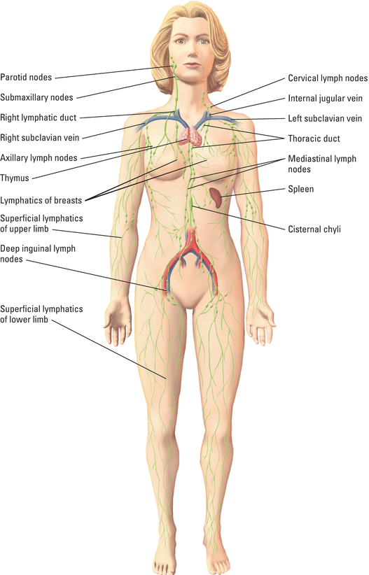
Drained by right lymph duct
Drained by thoracic duct
The lymphatic system begins peripherally, with lymph capillaries that absorb fluid. The capillaries proceed centrally to thin vascular vessels. These vessels empty into collecting ducts, which empty into major veins at the base of the neck.
