Acute surgical wounds are uncomplicated breaks in the skin that result from surgery. In an otherwise healthy individual, these types of wounds typically heal without incident. Surgical wounds heal by three methods:
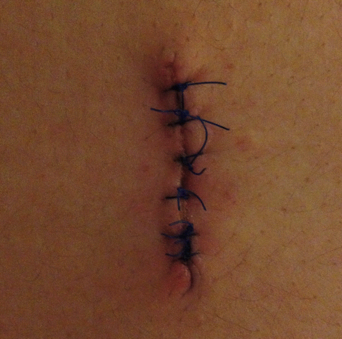
Primary intention: wound edges are approximated and secured.
Secondary intention: skin edges are not closed. Dressings are used to absorb drainage and promote healing. Used if infection is present. Wound heals by forming granulation tissue from the bottom out.
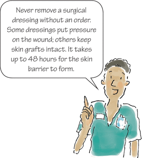
Third intention, or delayed primary closure, occurs when a contaminated wound is left open until contamination and inflammation resolve, and then closed by primary intention (this usually occurs after several days).
Assessment
First check the outside…
- Is the dressing stained?
- Estimate drainage quantity.
- Note its color, consistency, and odor.
- Estimate drainage quantity.
- Does the patient have a drainage device?
- Record the amount of drainage.
- Note the color of the drainage.
- Ensure that the device is patent, secure, and free from kinks.
- Record the amount of drainage.
- Does the patient have an ostomy (urostomy, colostomy, ileostomy)?
- Describe the output, color, location of the stoma, security of the pouching system, and peristomal skin.
- Describe the output, color, location of the stoma, security of the pouching system, and peristomal skin.
… then go under cover
- Is a healing ridge present?
- Are there signs and symptoms of infection, such as redness, warmth, and edema along the incision line and in the surrounding area; localized pain and tenderness; fever; pus or exudate from the incision; separation of suture line? Report these to the surgeon.
- Are there signs and symptoms of infection, such as redness, warmth, and edema along the incision line and in the surrounding area; localized pain and tenderness; fever; pus or exudate from the incision; separation of suture line? Report these to the surgeon.
Warning!
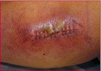
Wound infection is the most common surgical wound complication and the second most common infection type that occurs during hospitalization.
Postoperative leg wound infection
Definition: Healing ridge
“Palpable ridge that forms on each side of the wound during normal wound healing. It results from a buildup of collagen fibers, which begins to form during the inflammatory phase of wound healing and peaks during the proliferation phase (approximately 5 to 9 days postoperatively). Ridges typically fail to develop because of mechanical strain on the wound.”
| ||||||||||||||||||||||||||||
Wound closure
The severity and location of a wound determines the type of material used to close it. Newer technologies such as skin adhesives and negative pressure wound therapy designed for closed incisions are available to enhance healing.
Topical skin adhesives, such as Dermabond, are applied to the approximated incision and form a strong flexible bond to the skin. The film stays in place for 5 to 10 days. It is commonly used on facial lacerations and trocar puncture sites from laparoscopic procedures. Instruct the patient that he or she may shower and then pat the area dry gently. The patient should not swim, soak, or scrub the wound until the Dermabond falls off.
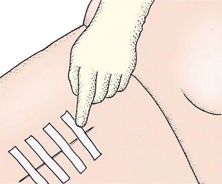
Adhesive closures, such as Steri-Strips or butterfly closures, may be used to close small wounds with scant drainage or to provide continued reinforcement after suture or staple removal. Apply 1/8″ apart to clean and dry skin. Do not apply under pressure to avoid blisters.
Steri-Strips are thin strips of sterile, nonwoven tape. They're a primary means of closing small wounds or holding a wound closed after suture/staple removal.
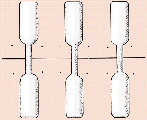
Butterfly closures consist of two sterile, waterproof adhesive strips linked by a narrow, nonadhesive “bridge.” They're used to hold small wounds closed after a laceration to promote healing after suture removal.
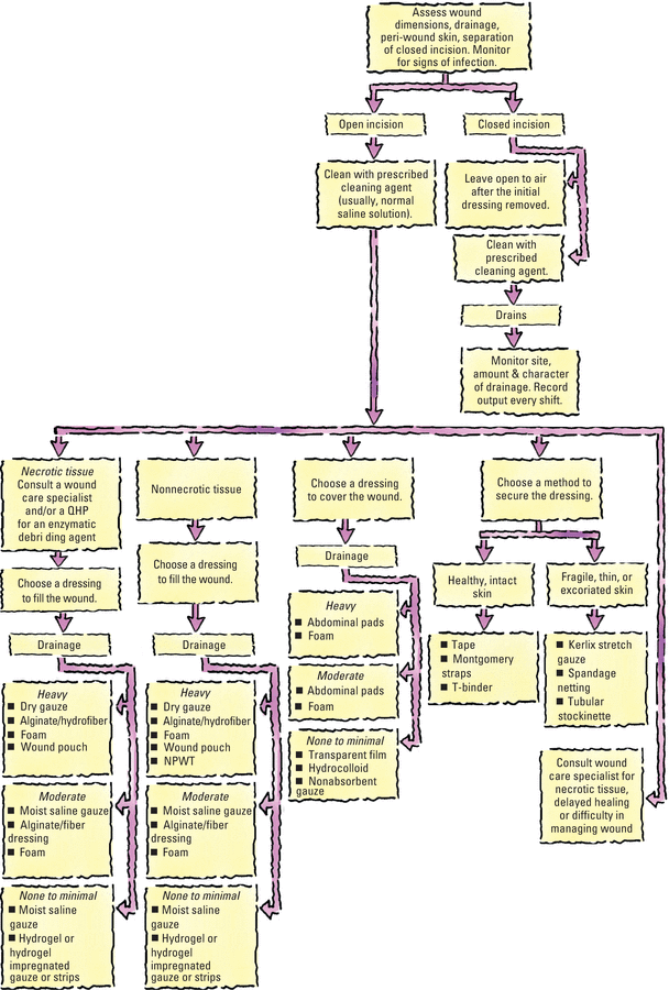
Surgeons usually use sutures. If cosmetic results aren't an issue, the surgeon may choose to use skin staples or clips.
- Used to close the skin surface
- Provide strength and immobility
- Cause minimal tissue irritation
- Consist of silk, cotton, stainless steel, or nylon.
- Broken down by the body so used when suture removal is undesirable
- Consist of:
- chromic catgut—a natural catgut treated with chromium trioxide to improve strength and prolong absorption time
- plain catgut—a material that's absorbed faster and is more likely to cause irritation than chromic catgut
- synthetic materials (such as polyglycolic acid)—materials that are replacing catgut because they're stronger, more durable, and less irritating.
- chromic catgut—a natural catgut treated with chromium trioxide to improve strength and prolong absorption time
An abdominal dressing requiring frequent changes can be secured with Montgomery straps to promote the patient's comfort and protect from MARSI (medical adhesive-related skin injury). If ready-made straps aren't available, follow these steps to make your own:
- Cut four to six strips of 2″ to 3″ wide hypoallergenic tape of sufficient length to allow the tape to extend about 6″ (15.2 cm) beyond the wound on each side. (The length of the tape might vary according to the patient's size and the type and amount of dressing.)
- Fold one of each strip 2″ to 3″ (5 to 7.5 cm) back on itself (sticky sides together) to form a nonadhesive tab. Then cut a small hole in the folded tab's center, close to its top edge. Make as many pairs of straps as you'll need to snugly secure the dressing.
- Clean the patient's skin to prevent irritation. After the skin dries, apply a skin protectant. Then apply the sticky side of each tape to a skin barrier wafer or a hydrocolloid wafer, and apply the wafer directly to the skin near the dressing.
- Thread a separate piece of gauze tie, umbilical tape, or twill tape (about 12″ [30.5 cm]) through each pair of holes in the straps, and fasten each tie as you would a shoelace. Don't stress the surrounding skin by securing the ties too tightly.
Replace Montgomery straps every 2 to 3 days or whenever they become soiled. If skin maceration occurs, place new tapes about 1″ (2.5 cm) away from irritation.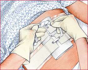
After surgery, bariatric patients experience slower wound healing because:
- Adipose tissue lacks a sufficient blood supply.
- The diaphragm doesn't descend completely, decreasing vital capacity.
- Insufficient oxygen slows digestion of bacteria by neutrophils.
They are also at increased risk for complications, such as:
- trauma (for example, the more forceful retraction needed during surgery may cause necrosis of the abdominal wall)
- infection (the difficulty level of operating on these patients lengthens the operation time, increasing the chances of contamination)
- dehiscence (excess fat and excess blood or serous fluid increase tension on the incision and wound's edges)
- hematoma.
- Assess the incision site and vital signs frequently.
- Use an abdominal binder over the surgical site to support the incision.
- Encourage the patient to use deep breathing and spirometry to improve oxygenation.
- Assess nutritional status and promote adequate intake of protein, carbohydrates, and vitamins.
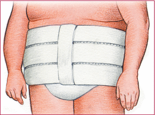
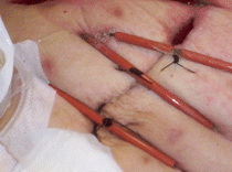
Although not used exclusively for bariatric patients, retention sutures are sometimes used after surgery in overweight patients to secure a wound's edges and reinforce the suture line. They can also be used for patients who have had multiple procedures in a short time or who are at high risk for wound dehiscence. Placed through the abdominal wall before the abdominal layers are closed, they provide support to deep tissue while the more superficial fascia and skin tissue heal. Monitor the entry site at the skin to prevent device-related pressure injuries. And keep the skin around these sutures clean.

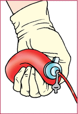
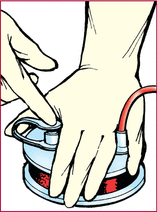
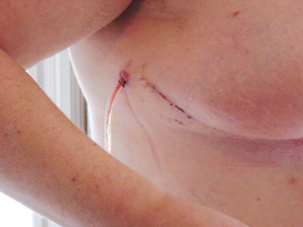
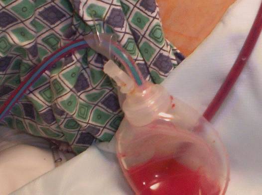
Surgeons insert closed wound drains during surgery when they expect a large amount of postoperative drainage. These drains suction serosanguineous fluid from the wound site.
If a wound produces heavy drainage, the closed wound drain may be left in place for longer than 1 week. Drainage must be frequently emptied and measured to maintain maximum suction and prevent strain on the suture line. Treat the tubing exit site as an additional surgical wound. Be sure to secure the drainage device so it does not pull or become dislodged.
- Promote healing
- Prevent swelling
- Reduce risk of infection and skin breakdown
- Minimize the need for dressing changes
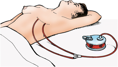
A closed wound drain consists of perforated tubing which facilitates drainage and is connected to a portable vacuum unit. (Hemovac and Jackson-Pratt are the most commonly used drainage systems.) The distal end of the tubing lies within the wound and usually leaves the body from a site other than the primary suture line. The drain is usually sutured to the skin. Shown below is a closed wound drainage system in a postmastectomy patient.
|
Surgical drain
Ostomy care
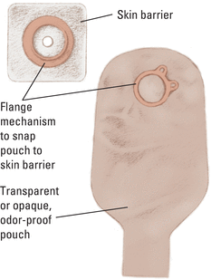
A patient with a urostomy, colostomy, or ileostomy wears an external pouch over the ostomy site, usually attached with a barrier wafer. The pouch collects urine or fecal matter, helps control odor, and protects the stoma and peristomal skin. Most disposable pouching systems can be used for 3 to 7 days, unless a leak develops.
When selecting a pouching system, choose one that delivers the best adhesive seal and skin protection for that patient. Other considerations include the stoma's location and structure, consistency of the effluent, availability and cost of supplies, amount of time the patient will wear the pouch, any known adhesive allergy, and the personal preferences of the patient.

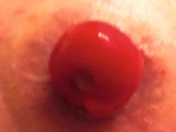
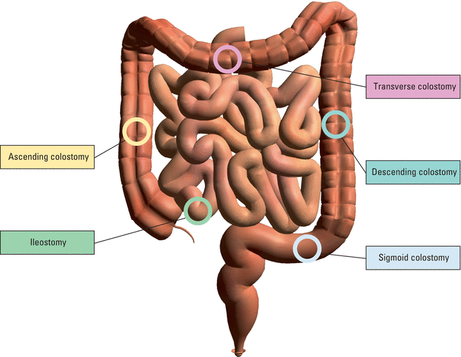
Comparing ostomy pouching systems
Manufactured in many shapes and sizes, ostomy pouches are fashioned for comfort, safety, and easy application. Some commonly available pouches are described here.
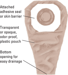
Patients who must empty their pouch often (because of diarrhea or a new colostomy or ileostomy) may prefer a one-piece, drainable, disposable pouch attached to a skin barrier. This pouch may be used permanently or temporarily, until stoma size stabilizes.
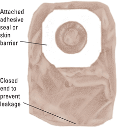
Disposable closed-end pouches, made of transparent or opaque odor-proof plastic, may come with a carbon filter for gas release. A patient with a regular bowel elimination pattern may choose this style for additional security and confidence.
A two-piece disposable drainable pouch with separate skin barrier permits frequent changes of the pouch while minimizing barrier removal.
Urostomy pouches have a spout on the end that can be connected to straight drainage for overnight or leg bag collection.
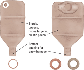
Reusable pouches come with a separate custom-made faceplate and O-ring (as shown at right). Some pouches have a pressure valve for releasing gas. The device has a 1- to 2-month life span, depending on how frequently the patient empties the pouch. It is secured to the abdomen with a belt.
Reusable equipment may benefit a patient who needs a firm faceplate, or has skin reactions to adhesives in the disposable barrier. However, many reusable ostomy pouches aren't as odor proof as the disposable pouches.
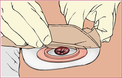
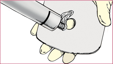
Fitting a skin barrier and ostomy pouch properly can be done in a few steps. Shown here is a two-piece pouching system with flanges, which is commonly used.
- Measure the stoma using a measuring guide.
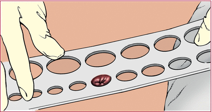
- Trace the appropriate circle carefully on the back of the skin barrier.
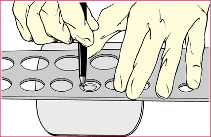
- Cut the circular opening in the skin barrier. Bevel the edges to keep them from irritating the patient. Be sure to size the opening correctly to prevent peristomal skin breakdown (approximately 1/8″ larger than the stoma).
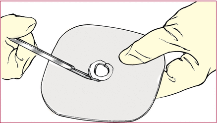
- Remove the backing from the skin barrier. Apply a barrier ring or paste, if needed, along the edge of the circular opening.
- Center the skin barrier over the stoma, adhesive side down, and gently press it to the skin.
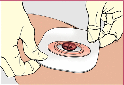
- Gently press the pouch opening onto the ring until it snaps into place.
