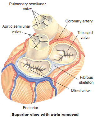
To prevent the backflow of blood during cardiac contraction, the atria and ventricles are separated from each other by two sets of valves composed of endocardial and connective tissue. The fibrous connective tissue prevents enlargement of valve openings and anchors valve flaps.
Atrioventricular Valves
The first set of heart valves, the atrioventricular (AV) valves, is located between each atrium and ventricle.
- Tricuspid valve-The right AV valve is between the right atrium and right ventricle. It derives its name “tricuspid” from its construction of three feathery leaflets, or cusps. Its function is to prevent backflow of blood from the right ventricle into the right atrium.
- Mitral valve-The left AV valve, commonly called the “mitral valve” is between the left atrium and left ventricle. The valve is known as a bicuspid valve since it only has two cusps. The mitral valve closes to keep blood from leaking backward when the left ventricle contracts.
The AV valves open to allow ventricular filling when the intra-atrial pressure exceeds the intraventricular pressure during atrial contraction. The onset of ventricular contraction creates pressure to close the AV valves.
Semilunar Valves
The other set of valves, called semilunar valves, allows blood to leave the heart via two major arteries, the aorta and pulmonary artery. They are one-way valves, meaning the blood cannot flow back into the heart after contraction. The two semilunar valves are the:
- Pulmonary valve-The pulmonary valve (sometimes referred to as the pulmonic valve) has three cusps and lies between the right ventricle and the pulmonary artery. The valve is opened by ventricular systole (contraction of the muscular tissue), pushing blood out of the right ventricle into the pulmonary artery.
- Aortic valve-The aortic valve is also a three-cusped valve located between the left ventricle and the aorta. With each of the heart muscle’s contractions, oxygenated blood exits the left ventricle through the aortic valve into the aorta
