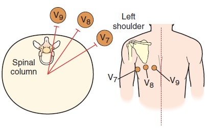
Areas of the heart that are not well visualized by the six chest leads include the wall of the right ventricle and the posterior wall of the left ventricle. A 15-lead ECG, which includes the standard 12 leads plus leads V7, V8, and V9, increases the chance of detecting an MI in these areas.
If a 15-lead cable is not available, use a standard 12-lead cable by repositioning leads V4, V5, and V6 to the back. They then become leads V7, V8, and V9 as shown below. Cross out the labels for V4, V5, and V6 and write in V7, V8, and V9 on the ECG paper.

The 15-Lead ECG
| Chest Leads | Electrode Placement | View of Heart |
|---|---|---|
| V7 | Level of V6 at posterior axillary line | Right ventricle |
| V8 | Aligned just below tip of scapula | Posterior wall of left ventricle |
| V9 | Directly between V8 and spinal column at posterior 5th intercostal space | Posterior wall of left ventricle |