Anatomy and Metabolism of Bone
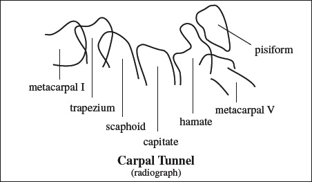
mnemonic:Some Lovers Try Positions That They Can't Handle
|
- Remember that trapezium comes before trapezoid in the dictionary as well!
- Spaces between carpal bones: ≤3 mm wide (internal comparison with capitolunate joint)
[scaphion, Greek = boat]
- The largest bone of proximal carpal row!
- Function: acts as an intercalated segment between lunate proximally + trapezium and trapezoid distally
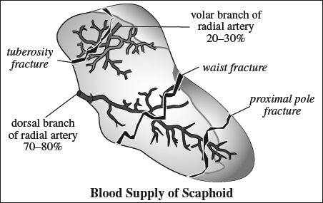
- Division: proximal third; middle third with waist; distal third with tuberosity on palmar surface
- Blood supply: branches of radial artery enter bone near midportion / waist dorsally → perfusion of mid to proximal 70–80% of scaphoid + proximal pole supplied by end artery in retrograde fashion
[luna, Latin = moon]
= moon-shaped configuration on LAT view
- Parts: body, volar pole, dorsal pole
- Function: acts as keystone of proximal carpal row
- Blood supply: single vessel (20%); 2 nonarticular nutrient arteries with consistent intraosseous anastomosis (80%)


- Blood vessels frequently enter distal half of bone putting proximal bone at risk for avascular necrosis!
|
= HULTEN VARIANCE = RADIOULNAR INDEX
= relative lengths of distal articular surfaces of radius and ulna
Definition:
- neutral = both surfaces at same level = equal length of ulna + radius
- positive = ulnar surface distal to radial surface = long ulna
- Gymnasts have a predilection for a positive ulnar variance!
Risk:- ulnar impaction (→ chondral fibrillation, chondromalacia, degenerative arthritis)
- ligamentous and carpal disturbances (→ TFCC tear, focal chondral lunate injury)
- negative = ulnar surface proximal to radial surface = short ulna
- Most children aged 12–16 years have a negative ulnar variance!
Effect of wrist position:
- increase of ulnar variance
- maximum forearm pronation
- firm grip
- decrease of ulnar variance
- maximum forearm supination
- cessation of grip
Radiographic standard view of unloaded wrist: posteroanterior, neutral forearm rotation, elbow flexed 90°, shoulder abducted 90°
[Jean Casimir Félix Guyon (1831–1920), 1st French chair in urology at University of Paris]
= canalis nervi ulnaris = loge de Guyon (French)
= small superficial tunnel-like structure at base of hypothenar
- Floor: depression between pisiform + hook of hamate
- Roof: volar carpal ligament and pisohamate ligament; retinaculum flexorum manus; flexor carpi ulnaris m.
- Contents: ulnar nerve with bifurcation into superficial and deep branches; ulnar artery
- Clinical significance: site for compression injury by anomalous muscle, ganglion, hamate fracture


Important Stabilizing Wrist Ligaments
- Important for carpal stability!
- Intrinsic = between carpal bones
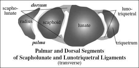
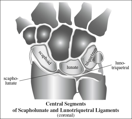
- Scapholunate lig.
- Lunotriquetral lig.
- Extrinsic = between carpal + metacarpal bones or between carpal bones + radius / ulna
- Palmar: carpal stability
- Radial collateral lig.
- Radiolunotriquetral lig. (RLTL): preventing ulnar translation
- Radioscaphocapitate lig. (RSCL): keeps scaphoid in position
- Dorsal radiocarpal ligg.: prevent perilunate instability + volar intercalated segment instability
- Dorsal intercarpal ligg.: prevent perilunate instability + dorsal intercalated segment instability
- Palmar: carpal stability