Anatomy and Metabolism of Bone
= quadriceps muscle consisting of
- Vastus medialis m.
- Vastus lateralis m.
- Vastus intermedius m.
- Rectus femoris m.
Insertion: combined as quadriceps tendon on patella
[pes, Latin = foot; anser, Latin = goose]
= tendinous configuration of 3 flexors + medial rotators of knee joint attaching inferomedially to tibial tuberosity
mnemonic:Say GraceSe before eating goose
- Sartorius tendon (anterior)
- Gracilis tendon (middle)
- Semitendinosus tendon (posterior)
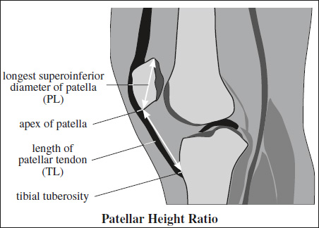
= high-riding patella as normal anatomic variant
- Cause: patellar tendon too long → reduced patellar contact area → patellar dislocation
- High-riding patella in 25% of patients with acute patellar dislocation
- mostly asymptomatic
MR measurement:
- patellar height ratio (Insall-Salvati index) = TL÷PL = patellar tendon length (TL) divided by superoinferior diameter of patella (PL) 1.1 ± 0.1 = normal; >1.3(M) or >1.5 (F) = patella alta; <0.74 (M) or <0.79 (F) = patella baja (infera)

- Both cruciate ligaments are intracapsular but extrasynovial!
Anterior Cruciate Ligament (ACL)
- Function: limits anterior tibial translation
- Origin: inner face of lateral femoral condyle
- Insertion: noncartilaginous region of anterior aspect of intercondylar eminence of tibia
- Anatomy: several distinct bundles of fibers
- large posterior bulk = spiraling together at femoral origin
- small anteromedial bundle diverging at tibial insertion
- thin solid taut dark band (sagittal MR with knee in extension) almost parallel to intercondylar roof (= Blumensaat line):
- with knee extension posterolateral band taut
- with increasing flexion:
anteromedial band becomes more taut + posterolateral band more lax
- thin hypointense band parallel to inner aspect of lateral femoral condyle + fanlike configuration toward tibial spine (coronal MR)
- thin ovoid hypointense band proximally, elliptical configuration distally with higher intensity (axial MR)
- greater SI than posterior cruciate ligament (due to anatomy)
Posterior Cruciate Ligament (PCL)
- Function: limits posterior tibial translation
- Origin: in a depression posterior to intercondylar region of tibia below joint surface
- Insertion: most distal + anterior aspect of inner face of medial femoral condyle
- thick dark band slightly posteriorly convex (arclike course on sagittal MR with knee in extension)
- medial to ACL (coronal MR)
Collateral Ligaments of Knee Joint
Medial (Tibial) Collateral Ligament
- Origin: just distal to adductor tubercle of femur
- Insertion: anteromedial face of tibia distal to level of tibial tubercle about 5 cm below joint line
- deep portion:
- meniscofemoral ligament
- meniscotibial ligaments
- superficial portion
- vertical band from femoral epicondyle to pes anserinus
- posterior oblique ligament = posterior oblique band from femoral epicondyle to semimembranosus tendon
- deep and superficial dark bands separated by a thin bursa + fatty tissue (on coronal MR)
Lateral (Fibular) Collateral Ligament
- Origin: lateral aspect of lateral femoral condyle
- Insertion: styloid process of fibular head
- bicipital tendon + iliotibial band join lateral collateral lig.
Arcuate Complex
- Function: provides posterolateral stabilization
- Consists of: lateral (fibular) collateral ligament + biceps femoris tendon + popliteus muscle and tendon + popliteal meniscal ligament + popliteal fibular ligament + oblique popliteal ligament + arcuate ligament + fabellofibular ligament + lateral gastrocnemius muscle
Posteromedial Corner of Knee
- Semimembranosus tendon
Attachment: infraglenoid tubercle of posteromedial tibia; posterior joint capsule; posterior horn of medial meniscus - Posterior joint capsule
- Posterior oblique ligament
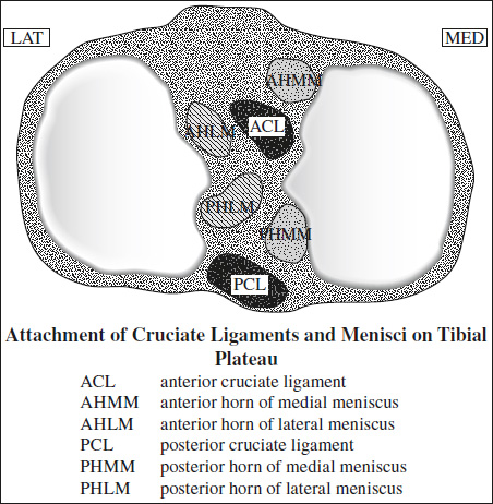
= wedge-shaped semilunar fibrocartilaginous structures
- Function: absorb shock, distribute axial load, assist in joint lubrication, facilitate nutrient distribution
- Margin:
- superior concave surface conforming to femoral condyle → increase in contact area
- inferior flat base that attaches to central tibial plateau via anterior + posterior root ligament anchors → maintain normal meniscal position + biomechanical function
- thick peripheral portion
- tapered central free edge
- Composition: collagen bundles oriented in
- circumferential (longitudinal) type I collagen bundles parallel to long axis of meniscus → hoop strength resisting axial load + preventing meniscal extrusion
- radial thin fibers perpendicular to longitudinal bundles forming a lattice → tying bundles together + providing structural support
- Subdivision into thirds:
- anterior horn
- meniscal body
- posterior horn
- Attachment:
- posterior root
- anterior root
- lateral meniscus (LM)
- striated / comb-like appearance of anterior horn ← intimate association between anterior root of lateral meniscus + ACL insertion site
- medial meniscus (MM)
- anomalous insertion paralleling ACL → mimicks MM tear
- anterior root may insert along anterior margin of tibia → mimicks MM subluxation

- peripheral attachment to deep fibers of medial collateral ligament → meniscus less mobile
- lateral meniscus (LM)
- Imaging:
- on sagittal images of menisci:
- “bow-tie” structure peripherally
- opposing triangles centrally
- posterior horn larger than anterior horn for MM
- anterior + posterior horns of similar size + shape for LM
- on coronal images:
- triangular shape through body of meniscus
- wedge-shaped through horn of meniscus
- on axial images:
- open C-shaped configuration of medial meniscus
- increase in width from anterior to posterior
- on sagittal images of menisci:
- Variants mimicking a tear:
- Transverse meniscal (geniculate) ligament (83–90%)
= thin fibrous band that connects and stabilizes the anterior horns of the menisci- overrides superior aspect of menisci before completely fusing to menisci
DDx: anterior root tear
Dx: Trace cross section of transverse ligament through infrapatellar fat pad on more central SAG images! - Meniscofemoral ligaments (MFL) in 89–93%
- Wrisberg ligament
- posterior to posterior cruciate ligament
- Humphrey ligament
- anterior to posterior cruciate ligament
mnemonic:under the hump (of the PCL)
- anterior to posterior cruciate ligament
Origin: superior + medial aspect of posterior horn of lateral meniscus
Insertion: lateral aspect of medial femoral condyle
Function: assist PCL + help control mobility of posterior horn of lateral meniscus during knee flexion + extension- demonstrated in ⅓ of cases on SAG images; usually limited to single most medial image!
- Wrisberg ligament
- Popliteomeniscal fascicles (visualized in 90%)
- anteroinferior fascicle = floor of popliteal hiatus
- posterosuperior fascicle = roof of popliteal hiatus
= synovial-lined fibrous bands that attach to posterior horn of lateral meniscus + help form popliteal hiatus
Function: stabilize posterior horn - Popliteal hiatus
- = separates lateral meniscus from joint capsule
- above posterior aspect of lateral meniscus on most superficial SAG slice!
- popliteal tendon moves behind + inferior to meniscus on adjacent deeper SAG sections!
- Oblique meniscomeniscal ligament (1–4%)
= connects meniscal horns in X-wise fashion on AXIAL image- medial oblique meniscomeniscal ligament
- anterior horn of medial meniscus to posterior horn of lateral meniscus
- lateral oblique meniscomeniscal ligament
- anterior horn of lateral meniscus to posterior horn of medial meniscus
- medial oblique meniscomeniscal ligament
- traverses intercondylar fossa between ACL and PCL
- Transverse meniscal (geniculate) ligament (83–90%)
Anatomic Variants of Menisci
- Diskoid meniscus
= abnormally shaped enlarged meniscus with further central extension onto tibial articular surface
Prevalence: 1.5–3% for lateral meniscus; 0.12–0.3% for medial meniscus
Side: lateral÷medial meniscus = 10÷1
Age: children, adolescents- body of meniscus measures ≥15 mm on midline coronal image
- ≥3 bow-tie shapes on contiguous sagittal (4-mm-thick) sections
- diffuse intrameniscal signal intensity ← increased meniscal vascularity
- Meniscal flounce
= rippled appearance of free nonanchored inner edge of medial meniscus
Prevalence: 0.2–0.3% of asymptomatic knees - Meniscal ossicle (rare)
Cause: developmental, degenerative, posttraumatic
Location: posterior horn of MM- calcification on radiograph mimicks loose body
- increased signal intensity can mimic a tear
- Chondrocalcinosis
Prevalence: 5– 15% (increasing with age)- calcifications on radiograph
- increased signal intensity → lowers sensitivity and specificity for detection of meniscal tear
Posterolateral Corner Structures
= arcuate complex
- Fibular collateral ligament
- Arcuate ligament
- Popliteus musculotendinous complex
- Popliteofibular ligament
- Fabellofibular ligament
- Posterolateral capsule
Popliteus Musculotendinous Complex
- Origin: posteromedial tibial surface proximal to soleal line forming floor of popliteal fossa
- Course: forms long strong popliteal tendon that passes underneath posterolateral joint capsule + arcuate ligament (extracapsular); enters knee through popliteal hiatus posteroinferiorly behind posterior horn of lateral meniscus; passes beneath lateral collateral ligament + tendon of biceps femoris
= intracapsular – extraarticular – extrasynovial - Attachment: popliteal notch on lateral aspect of lateral femoral condyle; anteroinferior to proximal attachment of lateral collateral ligament on lateral epicondyle
- Function: in non–weight-bearing state primary internal rotator of tibia on femur; in weight-bearing state external rotator of femur on leg
- fluid-filled popliteus bursa surrounds popliteus muscle and tendon
Fibular Collateral Ligament
= (TRUE) LATERAL COLLATERAL LIGAMENT
- Origin: lateral femoral epicondyle
- Attachment: lateral aspect of fibular head + neck anterior and distal to fibular styloid process; often conjoined insertion with biceps femoris tendon
- Function: simple passive restraint
- extracapsular WITHOUT meniscal attachment
Arcuate Ligament
= Y-shaped inconstant thickening of posterolateral capsule
- lateral limb inserts into posterolateral joint capsule
- medial limb extends medially over popliteus muscle to oblique popliteal ligament
Popliteofibular Ligament
= attachment of popliteus tendon to fibular head
- Origin: popliteus tendon near myotendinous junction
- Attachment: posterior aspect of fibular styloid process posteromedial to the biceps insertion
- Function: one of the strongest lateral stabilizers in knee
Fabella (present in 20%)
= sesamoid bone in lateral head of gastrocnemius muscle
- Function: anchors fabellofibular ligament
Fabellofibular ligament (present in 40%)
- Origin: fabella
- Attachment: styloid process of fibular head posterior to arcuate ligament + lateral to popliteofibular ligament / to lateral femoral condyle (in absence of fabella)
- Suprapatellar bursa
- Popliteal bursa
- Pes anserine bursa
- Semimembranosus-tibial collateral ligament bursa
- Prepatellar bursa
- Infrapatellar bursa
- Tibial collateral ligament bursa
- Tibiofibular joint cyst
- Popliteus bursa
- Meniscal cyst
- Cruciate ligament cyst
- Ganglion (mucinous degeneration of ACL)