Anatomy and Metabolism of Bone
Ligamentous Stabilizers of Ankle
Deltoid Ligament
= MEDIAL COLLATERAL LIGAMENT OF ANKLE
Function: main stabilizer against valgus force / pronation force / rotational force on talus
Components:
- superficial layer:
Origin: anterior colliculus of medial malleolus- Tibiocalcaneal ligament
Distal attachment: calcaneus - Tibionavicular ligament
Distal attachment: os naviculare - Posterior superficial tibiotalar ligament
Distal attachment: talus - Tibiospring ligament
Distal attachment: joins superomedial oblique band of spring lig. proper (= calcaneonavicular lig.)
Function: stabilizer for talocalcaneonavicular joint + medial plantar arch


- Tibiocalcaneal ligament
- deep layer = intraarticular, covered by synovium
Origin: intercollicular (= malleolar) groove + posterior colliculus of medial malleolus- Anterior tibiotalar ligament (ATTL)
- Posterior deep tibiotalar ligament (PDTL)
Deltoid ligament rupture is rare without additional injuries to the ankle ← uncommon occurrence of eversion ankle sprains and due to intrinsic thickness of the ligament.
Lateral Collateral Ligament Complex (LCL)
- 85% of all ankle sprains involve these ligaments!
- Anterior talofibular ligament (ATFL)
The anterior talofibular ligament is the weakest and most frequently injured among the 3 components of the lateral collateral ligament complex. - Posterior talofibular ligament (PTFL)
- Calcaneofibular ligament (CFL)
In inversion sprains, the calcaneofibular ligament is usually sequentially torn after the anterior talofibular ligament. If the anterior talofibular ligament is normal, then an isolated tear of the CFL is unlikely.
Syndesmotic Ligament Complex
binding tibia + fibula
- Anterior-inferior tibiofibular (AITFL)
Attachment: slightly above talofibular ligaments = above level of talotibial joint line- One of the most commonly injured ligaments in the ankle!
- Posteroinferior tibiofibular (PITFL)
- Transverse tibiofibular ligament
- Interosseous membrane
Anterior Tibialis
= most medial and largest extensor of ankle with appearance of tendon at junction of middle to distal ⅓
- Function: 80% of foot dorsiflexion; helps support longitudinal arch; aids in foot supination and inversion
- Origin: proximal third of lateral tibia, lateral tibial condyle, interosseous membrane, deep fascia, intermuscular septum
- Insertion: bifid = medial cuneiform (dominant slip) + 1st metatarsal base (thin slip)
- Tendon fixation: superior + inferior extensor retinaculum (with oblique superomedial and oblique inferomedial bands)
Extensor Hallucis Longus
- Function: extension of hallux
- Origin: middle half of fibula + interosseous membrane; becomes tendinous at distal ⅓ of tibia
- Insertion: dorsomedial surface of distal phalangeal base of hallux
- Course: descends vertically between anterior tibial and extensor digitorum longus muscles deep to superior + inferior extensor retinaculi
Extensor Digitorum Longus
- Function: extension of phalanges; contributes to foot dorsiflexion
- Origin: lateral tibial condyle, proximal ¾ of anterior fibula, interosseous membrane, deep fascia, intermuscular septa
- Insertion: dorsal aspect of middle + distal phalanges of 2nd through 5th digits
- Course: behind superior extensor retinaculum
Achilles Tendon
- Size: 7 mm in AP thickness (largest tendon of the body)
- Origin: gastrocnemius + soleus muscle
- surrounded by loose paratenon without tendon sheath
Plantaris Tendon
- parallels Achilles tendon anteromedially
- Insertion: Achilles tendon. calcaneus, plantar fascia
Posterior Tibialis Tendon
- Function: plantar flexion + inversion of foot; support for medial longitudinal arch of the foot
- Size: twice the size of flexor digitorum longus tendon
- Course: posterior + beneath medial malleolus (used as pulley) + flexor retinaculum; continues medial to subtalar joints
- largest anterior tendon component
- Insertion: navicular tuberosity + plantar capsule of navicular-cuneiform joint + plantar aspect of medial cuneiform bone
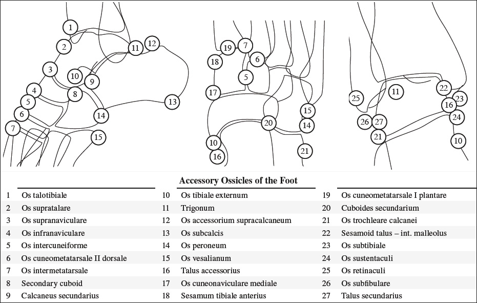
- Variation: ossified accessory navicular bone (in 25%) embedded into tendon
- deep middle (tarsometatarsal) tendon component
- Insertion: 2nd and 3rd cuneiform bones + cuboid bone + 2nd to 4th (± 5th) metatarsal bases

- posterior tendon component
- Origin: proximal tendon
- Insertion: anterior aspect of sustentaculum tali
Tendon sheath: ends 1–2 cm proximal to navicular insertion
Flexor Digitorum Longus Tendon
- Course: pierces medial intermuscular septum in medial-to-lateral direction; travels obliquely superficial (plantar) to flexor hallucis longus tendon; receives insertion of quadratus plantae muscle as it splits into four separate tendons giving rise to lumbrical muscles
- Insertion: bases of 2nd – 5th distal phalanges
Flexor Hallucis Longus Tendon
- Course: groove beneath posterior process of talus, beneath sustentaculum tali; pierces medial inter-muscular septum in medial-to-lateral direction; courses through fibro-osseous tunnel between tibial + fibular sesamoid bones of great toe
- Insertion: base of distal phalanx of hallux
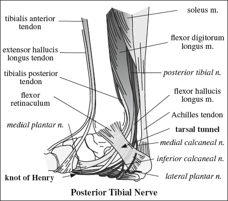
Flexor digitorum and hallucis longus tendons cross at the level of the navicular bone forming the master knot of Henry!
Peroneus Longus Tendon
The peroneus longus tendon is located posterolateral to peroneus brevis tendon in retromalleolar groove!
- Function: plantar flexion of 1st ray of foot + ankle and eversion of ankle; important stabilizer of ankle
- Insertion: fan-shaped onto lateral tubercle at base of 1st MT + plantar aspect of 1st cuneiform bone + base of 2nd MT bone + 1st dorsal interosseous muscle


- Course: posterior to lateral malleolus in retromalleolar groove; posterolateral to peroneus brevis tendon; beneath cuboid bone through cuboid fibro-osseous tunnel / groove; oblique anteromedial orientation while crossing plantar aspect of foot
- Variant: os peroneum (20%) = accessory ossicle embedded in peroneus longus tendon at level of calcaneocuboid joint
- N.B.: Tenosynovial effusion within plantar peroneus longus tendon sheath is uncommon
Fluid in plantar peroneus longus tendon sheath is highly suggestive of tenosynovitis!
Cysts and Bursae of the Ankle and Foot
- Malleolar bursa
- Retrocalcaneal bursa
- Tendo-Achilles bursa
- Tarsal tunnel ganglion
- Sinus tarsi bursa
- Intermetatarsal bursae
- medial compartment
- Border: medial septum (extending from plantar aponeurosis to navicular bone, medial cuneiform bone, plantar surface of 1st metatarsal bone)
- Content: abductor hallucis muscle + flexor hallucis brevis muscle + flexor hallucis longus tendon
- lateral compartment
- Border: lateral septum (extending from plantar aponeurosis to medial surface of 5th metatarsal bone)
- Content: abductor muscle + short flexor muscle + opponens muscle of 5th toe
- central compartment
- Border: medial + lateral septa; communicates directly with posterior compartment of calf; subdivided by horizontal septa: adductor hallucis muscle separated from quadratus plantae muscle
- Content: flexor digitorum brevis m. + flexor digitorum longus tendon + quadratus plantae muscle + lumbricales mm. + adductor hallucis muscle
- deep subcompartment
- Border: transverse fascia of forefoot; separated from quadratus plantae muscle
- Content: adductor hallucis muscle
= articulation between nine bones of forefoot and midfoot:
- 5 metatarsals (M1–M5) → contribute to plantar arch (M2–M4 bases of trapezoidal morphology at cross section and M2 base representing osseous “keystone”)
- 3 cuneiforms (C1–C3)
- cuboid
Three separate synovial articulations:
- lateral column = most mobile articulation of M4 + M5 with cuboid
- middle column = most rigid articulations of M2 + M3 with C2 + C3
- medial column = rigid articulation between C1 and M1
Location of Common Synovial Recesses and Bursae
|


= 2nd largest tarsal bone;
⅔of surface covered with articular cartilage
- head of talus
- Anterior articulation: Talonavicular joint
- Inferior articulation: Anterior talocalcaneal (subtalar) joint
- neck of talus
- Inferior surface: tarsal canal opening laterally into sinus tarsi
- talar body
- Inferior (subtalar) articulations:
- Posterior (lateral) facet: larger articulation
- Middle (medial) facet: sustentaculum
- Superior articulation: talar dome / trochlea
- tibiotalar joint
- Lateral articulation: fibula
- posterior facet of posterior talocalcaneal (subtalar) joint
- Inferior (subtalar) articulations:
- posterior process
- medial tubercle
- lateral tubercle
- elongated = Stieda process
- nonfused = os trigonum (prevalence of 2–50%)
- groove between both: for flexor hallucis longus tendon
Blood supply:
- Anterior tibial (dorsalis pedis) artery


- Posterior tibial artery → artery of tarsal canal
- Peroneal a. → peroneal perforating branch / lateral tarsal artery → tarsal sinus artery
Articulations:
- superiorly – subtalar joint:
- Anterior talar facet
- supported by calcaneal beak
- articulates with anterior talar facet
- Middle talar facet
- articulates with middle talar facet
- supported by sustentaculum tali
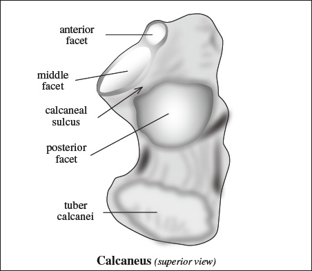
calcaneal sulcus = groove between middle and posterior facet
sinus tarsi = canal between calcaneal sulcus and talus - Posterior talar facet
thalamic portion = condensed cortical bone inferior to posterior facet
- Anterior talar facet
- anteriorly – calcaneocuboid joint
- triangular cuboidal facet
Lateral surface: flat + subcutaneous
- peroneal tubercle (centrally located)
- centrally
= attachment of calcaneofibular ligament centrally - anterosuperiorly
= attachment of lateral talocalcaneal ligament
- centrally

Medial surface:
- talar support by
- interosseous lig. + medial talocalcaneal lig.
- sustentaculum tali at anterior aspect of medial surface
- groove inferior to sustentaculum transmits tendon of flexor hallucis longus
- neurovascular bundle
