- 70% of blood flow is delivered to ICA
- shares waveform characteristics of both internal + external carotid arteries
- velocity increases toward the aorta (9 cm/sec for each cm of distance from the carotid bifurcation)
Carotid Bifurcation
= physiologic stenosis ← inertial forces of blood flow diverting main-flow stream from midvessel to a path along vessel margin at flow divider
Location: lateral to upper border of thyroid cartilage; at level of C3-4 intervertebral disk
Branches: ECA arises anterior + medial to ICA (95%)
External Carotid Artery Branches
mnemonic:All Summer Long Emily Ogled Peter's Sporty Isuzu
- Ascending pharyngeal artery
- Superior thyroid artery
- Lingual artery
- External maxillary = facial artery
- Occipital artery
- Posterior auricular artery
- Superficial temporal artery
- Internal maxillary artery
- CERVICAL SEGMENT
ascends posterior and medial to ECA; enters carotid canal of petrous bone; NO branches
Carotid bulb = carotid sinus:- = dilated proximal part of ICA with thinner media + thicker adventitia containing many receptor endings of glossopharyngeal nerve
- Function: baroreceptor responsive to changes in arterial blood pressure
- hypersensitive carotid sinus
= slight touch / head movement initiates
- vasodilatation with drop in blood pressure
- vagal stimulation with sinoatrial / atrioventricular cardiac block
- stagnant eddy that rotates at outer vessel margin
- PETROUS SEGMENT
ascends briefly in carotid canal → bends anteromedially in a horizontal course (anterior to tympanic cavity and cochlea); exits near petrous apex through posterior portion of foramen lacerum; ascends to juxtasellar location where it pierces dural layer of cavernous sinus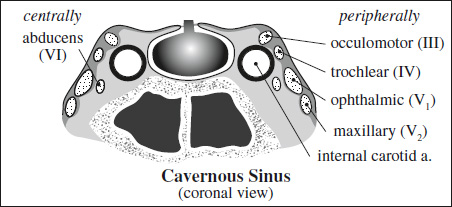
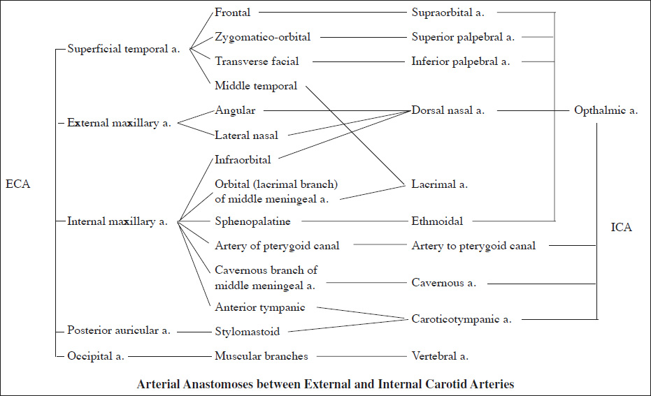
Branches:- Caroticotympanic a.: to tympanic cavity, anastomoses with anterior tympanic branch of maxillary a. + stylomastoid a.
- Pterygoid (vidian) a.: through pterygoid canal; anastomoses with recurrent branch of greater palatine a.
- CAVERNOUS SEGMENT ascends to posterior clinoid process → then turns anteriorly + superomedially through cavernous sinus; exits medial to anterior clinoid process piercing dura
Branches:- Meningohypophyseal trunk
- tentorial branch
- dorsal meningeal branch
- inferior hypophyseal branch
- Anterior meningeal a.: supplies dura of anterior fossa; anastomoses with meningeal branch of posterior ethmoidal a.
- Cavernous rami supply trigeminal ganglion, walls of cavernous + inferior petrosal sinuses
- Meningohypophyseal trunk
- SUPRACLINOID SEGMENT
ascends posterior + lateral between oculomotor + optic nerve
Branches:
mnemonic: OPA- Ophthalmic a.
- Posterior communicating a.
- Anterior choroidal a.
- Ophthalmic a. exits from ICA medial to anterior clinoid process, travels through optic canal inferolateral to optic nerve
- recurrent meningeal branch: dura of anterior middle cranial fossa
- posterior ethmoidal a.: supplies dura of planum sphenoidale
- anterior ethmoidal a.
- Superior hypophyseal a.: optic chiasm, anterior lobe of pituitary
- Posterior communicating a. (pCom)
- Anterior choroidal a.
- Middle + anterior cerebral arteries (MCA, ACA)
Flow direction: C4–C1
- C4 segment = before origin of ophthalmic a.

Duplex Identification of Carotid Arteries
Criteria External Carotid Artery Internal Carotid Artery Size usually smaller than ICA usually larger than ECA Location oriented medially + anteriorly toward face oriented laterally + posteriorly toward mastoid process (mnemonic: IAC vis-à-vis ECA positioned like helix vis-à-vis tragus of your ear) Branches gives off arterial branches (superior thyroid artery as 1st branch) NO arterial branches Waveform high-resistance flow pattern supplying capillary beds in skin + muscle: - forward systolic component
- early diastolic flow reversal, occasionally followed by another component
- little / no flow in late diastole
low-resistance waveform pattern supplying capillary bed in brain: - high-velocity forward systolic component
- sustained strong forward flow in diastole
- stagnant eddy with flow reversal opposite to flow divider in carotid bulb
Maneuver oscillations on temporal tap maneuver - C3 segment = genu of ICA
- C2 segment = supraclinoid segment after origin of ophthalmic a.
- C1 segment = terminal segment of ICA between pCom + ACA
Anterior Cerebral Artery (ACA)
A1 (horizontal) segment between origin and anterior communicating a. (aCom)
- inferior branches
supply superior surface of optic nerve + chiasm - superior branches
penetrate brain to supply anterior hypothalamus, septum pellucidum, anterior commissure, fornix columns, anterior inferior portion of corpus striatum - medial lenticulostriate artery (largest striatal artery) = recurrent artery of Heubner for anteroinferior portion of head of caudate, putamen, anterior limb of internal capsule
A2 (interhemispheric) segment after origin of anterior communicating a. (aCom); ascends in cistern of lamina terminalis
Branches:

- Medial orbitofrontal a.: along gyrus rectus
- Frontopolar a.
- Callosomarginal a.: within cingulus gyrus
- Pericallosal a.: over corpus callosum within callosal cistern
- Superior internal parietal a.: anterior portion of precuneus + convexity of superior parietal lobule
- Inferior internal parietal a.
- Posterior pericallosal a.
from callosomarginal / pericallosal artery:- anterior + middle + posterior internal frontal aa.
- paracentral a.: supplies pre- + postcentral gyri
Supply: anterior ⅔ of medial cerebral surface + 1 cm of superomedial brain over convexity
= largest branch of ICA arising lateral to optic chiasm
M1 (horizontal) segment = courses in lateral direction
Branches: lateral lenticulostriate aa.
Supply: part of head and body of caudate, globus pallidus, putamen, and posterior limb of internal capsule
M2 (sylvian) segment = enters sylvian fissure just ventral to anterior perforated substance; divides into superior and inferior divisions with 2 / 3 / 4 branches
Branches: temporal lobe and insular cortex (sensory language area of Wernicke), parietal lobe (sensory cortical areas), inferolateral frontal lobe
M3 (cortical) segment = distal branches lateral to insular cortex = candelabra [candelabrum, Latin = decorative candlestick / lamp with several arms or branches]
Branches:
- Anterior temporal artery
- Ascending frontal artery / prefrontal a.
- Precentral artery = pre-Rolandic a.
- Central artery = Rolandic a.
- Anterior parietal artery = post-Rolandic a.
- Posterior parietal artery
- Angular artery
- Middle temporal artery
- Posterior temporal artery
- Temporooccipital artery
Supply: lateral cerebrum, insula, anterior + lateral temporal lobe
originates from bifurcation of basilar artery within interpeduncular cistern (in 15% as a direct continuation of posterior communicating artery); lies above oculomotor nerve and circles midbrain above the tentorium cerebelli
Branches:
- Mesencephalic perforating branches: tectum + cerebral peduncles
- Posterior thalamoperforating aa.: midline of thalamus + hypothalamus
- Thalamogeniculate aa.: geniculate bodies + pulvinar
- Posterior medial choroidal a.: circles midbrain parallel to PCA; enters lateral aspect of quadrigeminal cistern; passes laterally and above pineal gland and enters roof of 3rd ventricle; supplies quadrigeminal plate + pineal gland
- Posterior lateral choroidal a.: courses laterally and enters choroidal fissure; anterior branch to temporal horn + posterior branch to choroid plexus of trigone and lateral ventricle + lateral geniculate body
- Cortical branches:
- Anterior inferior temporal artery
- Posterior inferior temporal artery
- Parietooccipital artery
- Calcarine artery
- Posterior pericallosal artery
Supply: medial + posterior temporal lobe, medial parietal lobe, occipital lobe
by multiple small vessels originating from Pcom + P1 and P2 segments of the PCAs
Territories:
- anterior: polar / thalamotuberal arteries ← Pcom
- paramedian: paramedian / thalamoperforating arteries ← P1 segment of PCA
- inferolateral: thalamogeniculate arteries ← P2 segment of PCA
- posterior: posterior choroidal arteries ← P2 segment
Anatomic variant (uncommon):
Artery of Percheron = single dominant thalamoperforating artery supplying both medial thalami (with variable contribution to rostral midbrain)
Occlusion:
- CHARACTERISTIC bilateral paramedian thalamic infarcts ± midbrain involvement
Arterial Anastomoses of the Brain
Anastomoses via Arteries at the Base of the Brain
- Circle of Willis
- Right ICA ↔ right ACA ↔ aCom ↔ left ACA ↔ left ICA
- ICA ↔ pCom ↔ basilar a.
- ICA ↔ anterior choroidal a. ↔ posterior choroidal a. ↔ PCA ↔ basilar a.
- Developmental anomaly four transient embryonal carotid-basilar anastomoses named according to their corresponding cranial nerves that regress in the following sequence:
- Primitive acoustic (otic) artery
= arterial connection between petrous portion of ICA within carotid canal + proximal basilar artery / posterior inferior cerebellar a.- traverses internal auditory canal (with CN VIII)
- Primitive hypoglossal artery
= arterial connection between the C1–C3 portion of ICA and proximal portion of basilar a.- traverses hypoglossal canal (with CN XII)
- Persistent primitive trigeminal artery
Frequency: ~ 1%- short wide connection between the cavernous portion of ICA + basilar artery (between anterior inferior cerebellar a. and superior cerebellar a.)
- penetrates sella turcica (in 50%)


- enlargement of ipsilateral ICA
- ectopic vessel crossing the pontine cistern to anastomose with basilar artery
- Proatlantal intersegmental artery
= arterial connection between CCA bifurcation / ECA (57%) / ICA at C2–C4 level (38%) and vertebral a. in suboccipital region- traverses foramen magnum
- Primitive acoustic (otic) artery
Anastomoses via Surface Vessels
- Leptomeningeal anastomoses of the cerebrum: ACA ↔ MCA ↔ PCA
- Leptomeningeal anastomoses of the cerebellum: Superior cerebellar a. ↔ AICA ↔ PICA
Rete Mirabile
ECA ↔ middle meningeal a. / superficial temporal a. ↔ leptomeningeal aa. ↔ ACA / MCA
Histo:NO smooth muscle / venous valves → bidirectional flow
Dural Venous Sinuses
= major drainage pathway from cerebral veins into internal jugular veins; enclosed by leaves of dura
- Superior group draining majority of brain + skull
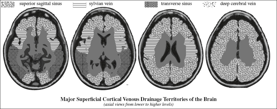
- Superior sagittal sinus (SSS) collects superficial cerebral veins that drain cerebral convexities
- luminal surface of triangular shape
- traversed by septa → maintaining laminar flow + preventing venous reflux into cortical veins
- Inferior sagittal sinus
- Straight sinus (SS)
- Occipital sinus small midline vein draining toward foramen magnum / into jugular fossa / suboccipital veins
Location: at attachment of falx cerebelli- may replace an aplastic transverse sinus
- Transverse sinus (TS)
receives blood from the temporal, parietal, and occipital lobes- commonly asymmetric with right dominance
- Sigmoid sinus
- Confluence of sinuses (torcular herophili) formed by union of the SSS +SS + TS
- often asymmetric in appearance (see below)
- Superior sagittal sinus (SSS) collects superficial cerebral veins that drain cerebral convexities
- inferior group drains superficial cerebral veins + basal and medial parts of undersurface of the brain + orbits + sphenoparietal sinus + cavernous sinus
- Cavernous sinus complex
- Superior petrosal sinus
- arising from junction of transverse with sigmoid sinus
- extends along petrous ridge
- Inferior petrosal sinus
- arising from distal portion of sigmoid sinus / jugular bulb
- extends along clivus
Variants of Torcular Herophili
- Codominant transverse sinuses (TS)
- Dominant right TS
- Dominant left TS
- Segmental hypo- / aplasia of proximal left TS + inflow to distal segment of TS from vein of Labbé and tentorial tributaries
- SSS drains into right TS + SS into left TS
- high split of SSS + SS drains into both TS
Superficial Venous System
= SUPERFICIAL CORTICAL VEINS
= great variability in drainage territories
- ascending (= superiorly draining) veins named for cortical area that is drained
- descending (inferiorly draining) veins
- Labbé vein
- Sylvian (superficial middle cerebral) vein drains blood from peri-insular region into basal dural sinuses
◊Relative luminal diameters of Trolard vein + Labbé vein + superficial sylvian vein are reciprocal
Location: subarachnoid space; traverse arachnoid mater + meningeal layer of dura mater to drain into dural venous sinus
Deep Venous System
= DEEP CEREBRAL VEINS
= centripetal drainage of hemispheric white matter and basal ganglia
- internal cerebral veins:
- Internal cerebral vein ← thalamostriate v. ← corpus callosum drainage (from anterior to posterior)
- septal vein
- anterior caudate vein
- terminal vein
- Basal vein of Rosenthal
- Vein of Galen
- Medullary + subependymal veins
- Internal cerebral vein ← thalamostriate v. ← corpus callosum drainage (from anterior to posterior)
- transcerebral veins: draining cerebral white matter
- typically not visualized due to small caliber
Infratentorial Venous System
- superiorly into vein of Galen
- anteriorly into petrosal sinuses
- posteriorly into dural sinuses
Important vascular markers:
- Pontomesencephalic v. = anterior border of brainstem
- Precentral cerebellar v. = position of tectum
- Colliculocentral point = midpoint of Twining's line at knee of precentral cerebellar vein
- Venous angle = acute angle at junction of thalamostriate with internal cerebral v. = posterior aspect of foramen of Monro
- Internal cerebral vv. = demarcate caudad border of splenium of corpus callosum superiorly + pineal gland inferiorly
- Copular point = junction of inferior + superior retrotonsillar tributaries draining cerebellar tonsils in region of copular pyramids of vermis
Arachnoid Granulations of Pacchioni
Prevalence: 66% (↑ in number + conspicuity with age)
Function: resorption of CSF
Location: within lacunae laterales of SSS
- well-defined 2–9 mm focal filling defects within dural sinus
- produce 13–15 mm calvarial impressions lateral to midline
- iso- (⅓) / hypoattenuating (⅔) relative to parenchyma


