Differential Diagnosis of Nervous System Disorders
Classification of Vascular CNS Anomalies
- VASCULAR MALFORMATION
- arterial = arteriovenous malformation (AVM)
- Classic brain AVM
- Cerebral proliferative angiopathy
- Cerebrofacial arteriovenous metameric syndrome
- Vein of Galen malformation
- capillary = capillary telangiectasia
- Capillary telangiectasia
- Facial port-wine stain
- venous = venous malformation
- Developmental venous anomaly
- Sinus pericranii
- lymphatic
- Cystic hygroma
- combinations
- Sturge-Weber disease
- Rendu-Osler-Weber disease
- arterial = arteriovenous malformation (AVM)
- VASCULAR TUMOR
- Hemangioma
- capillary hemangioma: seen in children, involution by 7 years of age in 95%
- cavernous hemangioma: seen in adults, no involution
- Hemangiopericytoma
- Hemangioendothelioma
- Angiosarcoma
- Hemangioma
= carotid (CA) + vertebral artery (VA) injuries during generalized multitrauma / direct craniocervical trauma
Prevalence: 1.1–1.6% of all blunt trauma
Mechanism: partial / complete failure of arterial mural integrity ← longitudinal stretching of artery, direct blow to artery, piercing by bone fragment
Prognosis: 25–38% mortality if injury untreated
Cx:
- Infarction ← intimal disruption / flap / hematoma
→ thromboembolism of platelet aggregates
→critical luminal stenosis + occlusion - Brain ischemia ← steal phenomenon by AV fistula
- Fatal exsanguination
Type of Arterial Injury
- Minimal intimal injury
- nonstenotic luminal irregularity
DDx: arterial spasm
- nonstenotic luminal irregularity
- Raised intimal flap
- linear intraluminal filling defect emanating from arterial wall
- Dissection with intramural hematoma
- eccentric / circumferential mural thickening:
- narrowed arterial lumen
- increased arterial diameter
- eccentric / circumferential mural thickening:
- Arterial occlusion
- lack of intraluminal enhancement
- Pseudoaneurysm
- eccentric outpouching from native arterial lumen:
- minimal contour abnormality
- large irregular saccular outpouching
- focal ballooning of arterial lumen
- eccentric outpouching from native arterial lumen:
- Transection with active hemorrhage
- irregular collection of extravascular contrast material surrounding parent vessel
- Arteriovenous fistula
- early venous enhancement during the arterial phase
- enlargement of draining vein
DDx:
- Atherosclerosis (presence of calcification, characteristic location, increasing age)
Location: vessel origin, carotid bulb, cavernous carotid segment - Coiled / looped cervical ICA segment (5–15%)
- Congenitally absent / small ICA (small / absent carotid canal)
Shunt Lesions of Cerebral Vasculature
- AV malformation
- AV fistula: pia, dura, carotid-cavernous sinus
- Vein of Galen malformation
- Developmental venous anomaly
- Cerebral cavernous malformation
- Sinus pericranii
- Embolic state:
- single vascular territory
- Hypoperfusive state:
- multiple vascular territories
Cause:
- Vasospasm from subarachnoid hemorrhage
- Embolic infarction (50%)
- thrombus (atrial fibrillation, valvular disease, atheromatous plaques of extracerebral arteries, fibromuscular dysplasia, intracranial aneurysm, surgery, paradoxic emboli, sickle cell disease, atherosclerosis, thrombotic thrombocytopenic purpura)
- fluctuating blood pressures; hypercoagulability
- cerebral petechial hemorrhage within cortical / basal gray matter during 2nd week (from fragments of embolus) in up to 40%; initial ischemia is followed by reperfusion (= HALLMARK of embolic infarction)
- “supernormal artery” on NECT = high-density material lodged in cerebral vessel near major bifurcations
- atheromatous narrowing of vessels
- fat
- nitrogen
- thrombus (atrial fibrillation, valvular disease, atheromatous plaques of extracerebral arteries, fibromuscular dysplasia, intracranial aneurysm, surgery, paradoxic emboli, sickle cell disease, atherosclerosis, thrombotic thrombocytopenic purpura)
- Watershed / border zone infarct (10%)
- Hypertension
- Hypertensive encephalopathy
- diffuse white matter hypodensity (edema ← arterial spasm)
- Hypertensive hemorrhage
Location: basal ganglia (putamen, external capsule), thalamus, pons, cerebellum - Lacunar infarction
- Subcortical arteriosclerotic encephalopathy
- Hypertensive encephalopathy
- Amyloidosis
involvement of small- + medium-sized arteries of meninges + cortex- normotensive patient >65 years of age
- multiple simultaneous / recurrent cortical hemorrhages
- Vasculitis
- Bacterial meningitis, TB, syphilis, fungus, virus, rickettsia
- Collagen-vascular disease: Wegener granulomatosis, polyarteritis nodosa, SLE, scleroderma, dermatomyositis
- Granulomatous angiitis: giant cell arteritis, sarcoidosis, Takayasu disease, temporal arteritis
- Inflammatory arteritis: rheumatoid arteritis, hypersensitivity arteritis, Behçet disease, lymphomatoid granulomatosis
- Drug-induced: IV amphetamine, ergot preparations, oral contraceptives
- Radiation arteritis = mineralizing microangiopathy
- Moyamoya disease
- Anoxic encephalopathy
cardiorespiratory arrest, near-drowning, drug overdose, CO poisoning - Venous thrombosis
Multiple Infarctions
◊Typical in extracranial occlusive disease, cardiac output problems, small vessel disease; in 6% from a shower of emboli
Location: usually bilateral + supratentorial (¾); supra- and infratentorial (¼)
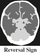
= inversion of the normal attenuation relationship between gray and white matter (gray matter of lower attenuation than adjacent white matter of thalami, brainstem, cerebellum) on NECT of brain
Pathogenesis: not fully understood
Cause: global cerebral injury with anoxic insult ← head trauma, nonaccidental trauma, hypoxia, drowning, status epilepticus, hypothermia, bacterial meningitis, strangulation
Prognosis: poor ← irreversible brain damage; survivors with profound neurologic deficits + severe developmental delay
Diffusion Weighted Imaging (DWI)
Hyperintense Lesion on DWI
- Cerebral infarction
- Epidermoid inclusion cyst
- Abscess with pus
- Encephalitis of cortex
- Creutzfeldt-Jakob disease
- Trauma: axonal shearing injury
- Neoplasm: medulloblastoma
Innumerable Punctate Hyperintense Lesions on DWI
= Starfield pattern
- Diffuse axonal injury (trauma)
- Emboli: cardiogenic, septic, fat
- Vasculitis
- Minute hemorrhagic metastases
Hypointense Lesion on DWI
- CSF
- Tumor cyst: pilocytic, hemangioblastoma
- Tumor nodules: hemangioblastoma
False Penumbra on Perfusion CT
True penumbra: successfully treatable with thrombolysis
- mean transit time (MTT) ↑
- cerebral blood flow (CBF) ↓
- cerebral blood volume (CBV) ↔
False penumbra: area of abnormal perfusion as in ischemic penumbra NOT treatable with thrombolysis
- Atherosclerosis at carotid bifurcation
= upstream flow limitation WITHOUT significant intracranial collateral blood supply- carotid bulb disease by CT angiography
- Evolving ischemic condition / chronic infarct
= delayed reperfusion + vascular collateralization of incomplete acute / chronic infarct- hypoattenuation in the same region
- restricted diffusion on DWI + ADC map
- Vascular dysregulation
= hyperemia on symptomatic side with apparent perfusion abnormality on contralateral normal side- clinical history!
Cause:- Hypertensive encephalopathy (PRES)
- perfusion abnormality ← vasospasm
- hyperintensity on STIR sequence
- no restricted diffusion
- Seizure, subarachnoid hemorrhage, hemiplegic migraine
- seizure activity
- no restricted diffusion
- Head tilt / angulation
- Variations in cerebrovascular anatomy
= physiologic perfusion delay in circle of Willis variants, unequal blood supply between anterior + posterior circulation, congenitally absent ICA, etc.
- LARGE-VESSEL VASCULITIS
- Takayasu arteritis
- Giant cell arteritis = Temporal arteritis
- MEDIUM-SIZED–VESSEL VASCULITIS
- Polyarteritis nodosa
- aneurysm / stenosis / occlusion of intracranial carotid arteries
- Kawasaki disease
- nonspecific subdural effusion, cerebral infarction
- reversible hyperintense lesion in splenium
- Polyarteritis nodosa
- SMALL-VESSEL VASCULITIS
- IgA vasculitis = Henoch-Schönlein purpura
- hypertensive encephalopathy
- focal ischemic / hemorrhagic lesions
- Microscopic polyangiitis
- cerebral hemorrhage + infarction, pachymeningitis
- variable degree of small-vessel disease
- Granulomatosis with polyangiitis
= Wegener granulomatosis- leptomeningeal enhancement
- Eosinophilic granulomatosis with polyangiitis
= Churg-Strauss syndrome- macro- / microinfarctions + macro- / microhemorrhages
- optic neuropathy
- IgA vasculitis = Henoch-Schönlein purpura
- VARIABLE-SIZED VESSEL VASCULITIS
- Behçet disease
- Cogan syndrome
- nonspecific ischemic changes
- obliteration / narrowing of vestibular labyrinth
- SINGLE-ORGAN VASCULITIS
- Primary angiitis of CNS (PACNS)
- discrete / diffuse supra- and infratentorial lesions
- ± areas of infarction and hemorrhage
- Primary angiitis of CNS (PACNS)
- VASCULITIS OF SYSTEMIC DISEASE
- Systemic lupus erythematosus (SLE)
- subcortical + periventricular white matter hyperintensity (60%)
- cerebral atrophy (30%), intracranial hemorrhage (3%)
- Sjögren syndrome
- extensive white + gray matter lesions + microbleeds
- Rheumatoid arthritis
- pachymeningitis with leptomeningeal enhancement
- cerebral vasculitis (rare)
- APLA (antiphospholipid antibody) syndrome
- arterial / venous thrombosis, thrombocytopenia
- Scleroderma
- nonspecific infarction, macro- and microhemorrhages
- Systemic lupus erythematosus (SLE)
- VASCULITIS WITH PROBABLE ETIOLOGY
- Infection-induced vasculitis
- Acute septic meningitis
- cerebral infarcts (5–15% in adults, 30% in neonates)
- Mycobacterium tuberculosis
- vasculitis of smaller cerebral arteries → small infarcts in basal ganglia
- Neurosyphilis
- predominantly MCA stroke in young adult
- Viral (in childhood):
- HIV-related vasculitis
- aneurysm, vessel occlusion, embolic disease, venous thrombosis
- Varicella-zoster vasculopathy
- uni- / bilateral basal ganglia infarcts
- HIV-related vasculitis
- Fungal: mucormycosis aspergillosis
- Parasitic: cysticercosis
- Malignancy-induced vasculitis
- Drug-induced vasculitis
- cocaine
- vasculitis, vasospasm, infarction, moyamoya-like
- heroin
- spongiform leukoencephalopathy
- cocaine
- Radiation-induced vasculitis
- wall thickening + prominent wall enhancement in affected large arteries
- Reversible cerebral vasoconstriction syndrome: Call-Fleming syndrome, postpartum angiopathy, migrainous vasospasm, benign angiopathy of CNS, vasoactive substances (cannabis, selective serotonin recapture inhibitors, nasal decongestants)
Imaging signs of cerebral vasculitis:
- direct
- vessel wall thickening
- vessel wall enhancement with contrast material
- indirect
- cerebral perfusion deficit
- ischemic brain lesion
- intracerebral / subarachnoid hemorrhage
- vascular stenosis