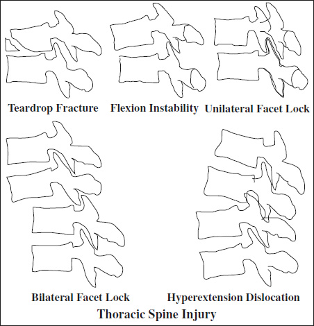
◊40% of all vertebral fractures that cause neurologic deficit; mostly complex (body + posterior elements involved)
Location:⅔ at thoracolumbar junction
Morphology:
- Compression
= loss of vertebral body height / disruption of vertebral endplate- vertebral height loss (approximate percentage!)
- degree of kyphosis
- Burst
= compression of posterior vertebral body + varying degrees of retropulsion- “burst” fragments at superior surface of body
- retropulsion of body fragments into spinal canal:
- = distance of line drawn between posterior margins of adjacent vertebral bodies + most posterior margin of bone fragment
- narrowing of spinal canal (approximate percentage!)
- Translation / rotation
= horizontal displacement or rotation of one vertebral body with respect to another- rotation of spinous processes
- uni- / bilateral facet fracture-dislocation
- vertebral subluxation
- Distraction
= dissociation along vertical axis ← disruption of anterior and posterior ligaments + osseous elements- diastasis of apophyseal joints:
- widening of facet joints
- empty “naked” facet joints
- perched / dislocated facet joints
- widening of interspinous space
- avulsion fracture of superior / inferior aspects of contiguous spinous processes
- vertebral body translation / rotation
Cx: kyphotic progression → subsequent vertebral collapse
Injury of Posterior Ligament Complex
MRI (only modality for direct assessment!):
- disruption of hypointense black stripe on sagittal T1WI / T2WI = tear of supraspinous ligament / ligamentum flavum / interspinous ligament
- fluid in facet capsules
- interruption of disk
- tear of anterior / posterior longitudinal ligaments
- edema in interspinous region = capsular / interspinous ligament injury
CT:
- widening of facet joints
- widening of interspinous distance
- spinous process avulsion fracture
- significant vertebral body subluxation / dislocation /translation
- UNRELIABLE: loss of vertebral body height, kyphosis
N.B.: inverse relationship between osseous destruction and ligamentous injury!
Fracture of Upper Thoracic Spine (T1 to T10)
Frequency: in 3% of all blunt chest trauma
Types:
- Compression / axial loading fracture (most common)
- wedging of vertebral body
- retropulsion of bone fragments
- posttraumatic disk herniation

- Burst fracture (more severe compression fracture)
- associated fracture of posterior neural arch
- comminuted retropulsed bone fragments
- Sagittal slice fracture
- vertebra above telescopes into vertebra below, displacing it laterally
- Anterior / posterior dislocation
- torn anterior / posterior longitudinal ligament
- facet dislocation
- Relatively stable fractures due to rib cage + strong costovertebral ligaments + more horizontal orientation of facet joints!
- Only 51% detected on initial CXR!
Often associated with: fracture of sternum
- widening of paraspinal lines
- mediastinal widening
- loss of height of vertebral body
- obscuration of pedicle
- left apical cap
- deviation of nasogastric tube
Signs of Spinal Instability:
= inability to maintain normal associations between vertebral segments while under physiologic load
- displaced vertebra
- widening of interspinous / interlaminar distance
- facet dislocation
- disruption of posterior vertebral body line
Fracture of Thoracolumbar Junction (T11 to L2)
= area of transition between a stiff + mobile segment of spine
- neurologic deficit (in up to 40%)
Classification based on injury to the middle column:
- Hyperflexion injury (most common)
= compression of anterior column + distraction of posterior spinal elements- hyperflexion-compression fracture
- loss of height of vertebral body anteriorly + laterally
- focal kyphosis / scoliosis
- fracture of anterosuperior endplate
- flexion-rotation injury (unusual)
- Very unstable!
- catastrophic neurologic sequelae: paraplegia
- subluxation / dislocation
- widening of interspinous distance
- fractures of lamina, transverse process, facets, adjacent ribs
- shearing fracture-dislocation
= damage of all 3 columns ← horizontally impacting force - flexion-distraction injury: Chance fracture
- hyperflexion-compression fracture
- Hyperextension injury (extremely uncommon)
- widened disk space anteriorly
- posterior subluxation
- vertebral anterior superior corner avulsion
- posterior arch fracture
- Axial compression fracture
- Unstable!
- burst fracture with herniation of intervertebral disk through endplates + comminution of vertebral body
- marked anterior vertebral body wedging
- retropulsed bone fragment
- increase in interpediculate distance
- ± vertical fracture through vertebral body, pedicle, lamina
= SEATBELT FRACTURE
[George Quentin Chance, British radiologist in Manchester, England]
Mechanism: shearing flexion-distraction injury (lap-type seatbelt injury in back-seat passengers)
- neurologic deficit infrequent (20%)
Location: L2 or L3
- horizontal splitting of spinous process, pedicles, laminae + superior portion of vertebral body
- disruption of ligaments
- distraction of intervertebral disk + facet joints
- Fracture often unstable!
Often associated with:
- other bone injury
rib fractures along the course of diagonal strap; sternal fractures; clavicular fractures - soft-tissue injury
transverse tear of rectus abdominis muscle; anterior peritoneal tear; diaphragmatic rupture - vascular injury
mesenteric vascular tear; transection of common carotid artery; injury to internal carotid artery, subclavian artery, superior vena cava; thoracic aortic tear; abdominal aortic transection - visceral injury
perforation of jejunum + ileum >large intestine >duodenum (free intraperitoneal fluid in 100%, mesenteric infiltration in 88%, thickened bowel wall in 75%, extraluminal air in 56%); laceration / rupture of liver, spleen, kidneys, pancreas, distended urinary bladder; uterine injury
Chance Equivalent
= purely ligamentous disruption leading to lumbar subluxation / dislocation
- mild widening of posterior aspect of affected disk space
- widened facet joints
- splaying of spinous processes = “empty hole” sign on AP view
[Sir Frank Wild Holdsworth (1904–1969), British pioneering orthopedist in rehabilitation of spinal injuries]
Location: thoracolumbar junction
- unstable spinal column fracture-dislocation with fracture through vertebral body + articular processes
- rupture of posterior spinal ligaments
= injury caused by three-point restraint type (combined lap and shoulder belt device)
- bruise in subcutaneous tissue + fat of anterior chest wall
- skin abrasions are associated with significant internal injuries (in 30%)
- Skeleton
sternum, ribs (along diagonal course of shoulder harness), clavicle, transverse processes of C7 or T1 - Cardiovascular
aortic transection, cardiac contusion, ventricular rupture, subclavian artery, SVC - Airways
tracheal / laryngeal tear, diaphragmatic rupture
- Skeleton
Transverse Process Fracture of Lumbar Spine
Cause: direct trauma, violent lateral flexion-extension forces, avulsion of psoas muscle, Malgaigne fracture
Frequency: 7%
In 21–51% associated injury:
- genitourinary injury, hepatic + splenic laceration
Location: L3 >L2 >L1 >L4 >L5; L÷R = 2÷1; multiple÷single = 2÷1; unilateral÷bilateral = 20÷1
- vertical÷horizontal (94%÷6%) fractures
- associated lumbar burst / compression fracture
- Detection by conventional radiography in 40% only!
Prognosis: minor and stable injury; 10% mortality
- Zone 1 = fracture lateral to sacral foramina
- significant neurologic deficit (uncommon)
- Zone 2 = fracture through ≥1 foramina
- unilateral lumbar / sacral radiculopathy (rare)
- Zone 3 = fracture through central canal
- significant bilateral neurologic damage (frequent): bowel / bladder incontinence
Cx: chronic disability (in up to 50%)
Acute Atraumatic Compression Fractures of Spine
Osteoporotic Compression Fracture of Vertebra
- low-signal-intensity band on T1WI and T2WI (93% sensitive)
- spared normal bone marrow SI of the vertebral body (85% sensitive)
- retropulsion of a posterior bone fragment into spinal canal (60% sensitive)
- “fluid” sign = circumscribed fluidlike SI on T2WI + STIR subjacent to fractured end plate
- multiple compression fractures
- rimlike enhancement around low-signal-intensity bands
- “wafer-like” distribution of radionuclide activity along endplate
Metastatic Compression Fracture of Vertebra
- convex posterior border of vertebral body (74% sensitive)
- abnormal SI of the pedicle or posterior element on enhanced fat-suppressed T1WI
- epidural mass
- encasing epidural mass
- focal paraspinal soft-tissue mass (41% sensitive)
- other sites of spinal metastases without compression fractures (63% sensitive)
- enhancement of metastatic foci
- completely replaced bone marrow of vertebral body (by tumor cells before trabeculae critically weakened)
Cx: spinal cord compression