Anatomy of Skull and Spine
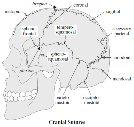
- METOPIC / FRONTAL SUTURE

= from nasion to anterior angle of bregma
[bregma, Greek = top of head]
Closure: by 3 months – 6 years of age; in up to 10% open until adulthood
Sutura frontalis persistens = metopism
= no closure of incomplete / complete metopic suture
DDx: anterior vertical fracture - SAGITTAL SUTURE
[sagitta, Latin = arrow]
= fibrous connective tissue joint between two parietal bones
Average width: 5.0 ± 0.2 mm (at birth), 2.4 ± 0.1 mm (1 month of age); narrowing further over time
Closure: 21–30 years of age; fusing anteriorly beginning at intersection with lambdoid suture - CORONAL SUTURE
= separates frontal from parietal bones
Average width: 2.5 ± 0.1 mm (at birth), 1.3 ± 0.1 mm (1 month of age)
Closure: 24 years of age - SQUAMOSAL SUTURE
- temporosquamosal suture
= connects temporal bone squama with lower border of parietal bone; arches posteriorly from pterion (= contact point between frontal, parietal, temporal, sphenoid)
[pteron, Greek = wing]
- often visualized at two points at CT with lambdoid suture acting as a useful posterior reference point
= continuous posteriorly with parietomastoid suture uniting mastoid process of temporal bone with region of mastoid angle of parietal bone - sphenosquamosal suture
= courses inferiorly from pterion separating sphenoid bone from squama of temporal bone
N.B.: often mistaken for skull base fracture
- LAMBDOID SUTURE
[upper case Greek letter lambda = L]
= connects parietal with occipital bone
Closure: 26 years of age
N.B.: the most common site of wormian bones - OCCIPITOMASTOID SUTURE

= inferior continuation of lambdoid suture at the point where lambdoid suture intersects with temporosquamosal suture - PARIETOMASTOID SUTURE
= links temporosquamosal and lambdoid sutures
- often not seen on axial CT images
- OCCIPITOMASTOID SUTURE
= between occipital bone + mastoid process of temporal bone as a continuation of the lambdoid suture toward skull base
N.B.: not infrequently mistaken for a skull base fracture - SPHENOFRONTAL SUTURE
= transverse suture between anterior margin of lesser sphenoid wing + posterior margin of horizontal orbital plate
- lesser sphenoid wing (posterior to suture) is a useful landmark for suture localization
- ACCESSORY PARIETAL SUTURE (rare)
= the most common of all usually bilateral and symmetric accessory sutures
Location: parietal and occipital bone → multiple ossification centers - MENDOSAL / ACCESSORY OCCIPITAL SUTURE
Frequency: 3% in an Indian subcontinent population
Closure: in utero / first few days of life; may persist up to 6 years of age
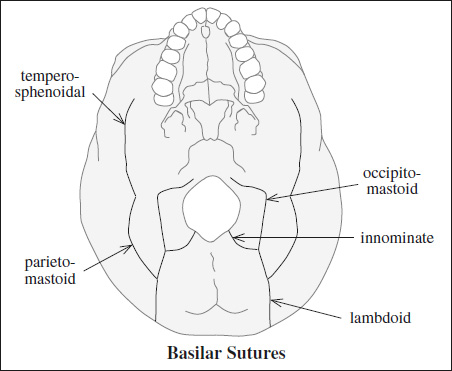
Os incae = large single centrally located intrasutural bone at junction of lambdoid and sagittal sutures; often forms in a persistent mendosal suture - SKULL BASE SUTURES
Ossification: 50% (84%) of anterior base by 6 (24) months
- innominate / intraoccipital
Closure: 4 years of age - lambdoid
- occipitomastoid
- parietomastoid
- temporosphenoidal
Symmetry and knowledge of the anatomic appearances of basal sutures are important for avoiding misdiagnosis.
A persistent hypoattenuating area of any length extending from foramen magnum beyond 4 years of age indicates a fracture.

