Motion mechanics: flexion, extension, minimal rotation
Physiologic range of rotation: 25°–53°
Major joint stabilizers (thickest + strongest ligaments):
- transverse lig. → allowing axial rotation
- alar ligg. → restrictors of lateral flexion, limiting excessive rotation
- joint capsules
- INTRINSIC LIGAMENTS (= 3 layers anterior to dura mater)
- Odontoid ligament
- Apical ligament
Function: secondary stabilizer preventing anterior shift
Course: from middle aspect of tip of odontoid process → anterior margin of foramen magnum - Alar ligaments (paired)
Function: secondary stabilizer preventing anterior shift
Course: from lateral aspect of tip of dens → medial aspect of occipital condyles
- Apical ligament
- Cruciate (cross-shaped) ligament
- Transverse ligament of atlas
Function: primary stabilizer of joint preventing excessive anterior motion of atlas on axis
Course: between medial portions of lateral masses: horizontal course behind dens - Crus superioris
Location: superior extension from transverse lig.
Attachment: lower margin of occipital bone - Crus inferioris
Location: inferior extension from transverse lig.
Attachment: posterior surface of body of axis
- Transverse ligament of atlas
- Tectorial membrane
= rostral continuation of posterior longitudinal lig.
Course: from body of C2 → anterior margin of foramen magnum
Function: restricts extension
- Odontoid ligament
- EXTRINSIC LIGAMENTS
= fibroelastic membranes as rostral continuations of- Anterior longitudinal lig.
- Ligamentum flavum
- Nuchal ligament
= continuation of inter- and supraspinous ligaments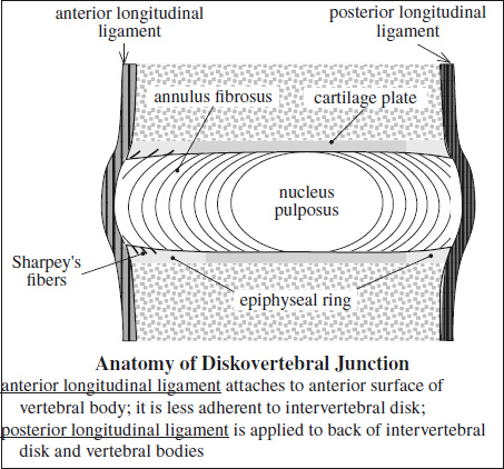
Course: 7th cervical vertebra → external occipital protuberance
Function: restriction of hyperflexion - Posterior atlanto-occipital membrane
= cephalic projection of ligamentum flavum
Course: posterior arch of atlas → posterior margin of foramen magnum