Differential Diagnosis of Skull and Spine Disorders
- POSTERIOR ARCH ANOMALIES
- Posterior atlas arch rachischisis (4%)
location: midline (97%); lateral through sulcus of vertebral artery (3%)- absence of arch-canal line (LAT view)
- superimposed on odontoid process / axis body simulating a fracture (open-mouth odontoid view)
- Total aplasia of posterior atlas arch
- Keller-type aplasia with persistence of posterior tubercle
- Aplasia with uni- / bilateral remnant + midline rachischisis
- Partial / total hemiaplasia of posterior arch
- Posterior atlas arch rachischisis (4%)
- ANTERIOR ARCH ANOMALIES
- Isolated anterior arch rachischisis (0.1%)
- Split atlas = anterior + posterior arch rachischisis
- plump rounded anterior arch overlapping the odontoid process making identification of predental space impossible (LAT view)
- duplicated anterior margins (LAT view)

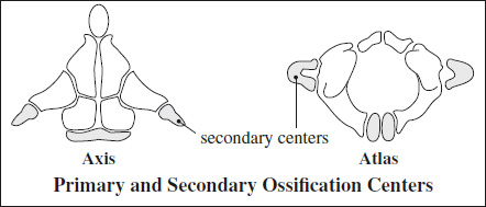
- Persistent ossiculum terminale = Bergman ossicle
- unfused odontoid process >12 years of age
DDx: type 1 odontoid fracture
- unfused odontoid process >12 years of age
- Odontoid aplasia (extremely rare)
- Os odontoideum
= independent os cephalad to axis body in location of odontoid process- absence of odontoid process
- anterior arch of atlas hypertrophic + situated too far posterior in relation to axis body
Cx: atlantoaxial instability
DDx: type 2 odontoid fracture (uncorticated margin)
Odontoid Erosion
mnemonic: P LARD
- Psoriasis
- Lupus erythematosus
- Ankylosing spondylitis
- Rheumatoid arthritis
- Down syndrome
= displacement of atlas with respect to axis
- Posterior atlantoaxial subluxation (rare)
- Anterior atlantoaxial subluxation (common)
= distance between dens + anterior arch of C1 (measurement along midplane of atlas on lateral view):- predental space: >2.5 mm >4.5 mm (in children)
- retrodental space: <18 mm
Causes of subluxation:
- Congenital
- Occipitalization of atlas 0.75% of population; fusion of basion + anterior arch of atlas
- Congenital insufficiency of transverse ligament
- Os odontoideum / aplasia of dens
- Down syndrome (20%)
- Morquio syndrome
- Bone dysplasia
- ARTHRITIS
due to laxity of transverse ligament or erosion of dens- Rheumatoid arthritis
- Psoriatic arthritis
- Reiter syndrome
- Ankylosing spondylitis
- SLE
rare: in gout + CPPD
- INFLAMMATORY PROCESS
pharyngeal infection in childhood, retropharyngeal abscess, coryza, otitis media, mastoiditis, cervical adenitis, parotitis, alveolar abscess- dislocation 8–10 days after onset of symptoms
- TRAUMA (very rare without odontoid fracture)
- MARFAN DISEASE
mnemonic: JAP LARD- Juvenile rheumatoid arthritis
- Ankylosing spondylitis
- Psoriatic arthritis
- Lupus erythematosus
- Accident (trauma)
- Retropharyngeal abscess, Rheumatoid arthritis
- Down syndrome
Pseudosubluxation of Cervical Spine
= ligamentous laxity in infants allows for movement of the vertebral bodies on each other, esp. C2 on C3
