Differential Diagnosis of Musculoskeletal Disorders
- Is there a lesion?
- Is it a bone tumor?
- Is the tumor benign or malignant?
- Is a biopsy necessary?
- Is histologic diagnosis consistent with radiographic image?
Assessment of Bone Tumor
A systematic approach is imperative for assessment of a bone tumor with attention to size, number, and location of lesions; margins and zone of transition; periosteal reaction; matrix mineralization; soft-tissue component.
- Age (and gender) of patient
- Precise tumor location
- transverse: medullary, cortical, juxtacortical
- longitudinal: epi-, meta-, diaphyseal
- Pattern of bone destruction / aggressiveness
- nonaggressive
- well-defined sharp margins
- smooth solid-appearing periosteal reaction
- aggressive infiltrative osseous process
- broad zone of transition
- poorly defined borders
- disrupted / “sunburst” appearance
- DDx: destructive metabolic / infectious process
- nonaggressive
- Lesion matrix
- “rings-and-arcs” appearance = chondral origin
- opaque cloud-like matrix = osseous mineralization
- osteolytic lesion → FEGNOMASHIC
- CT for cortical continuity / disruption
Action Following Bone Tumor Assessment
- BENIGN
- Diagnosis certain: no further work-up necessary
- Asymptomatic lesion with highly probable benign diagnosis may be followed clinically
- Symptomatic lesion with highly probable benign diagnosis may be treated without further work-up
- CONFUSING LESION
not clearly categorized as benign or malignant; needs staging work-up - MALIGNANT: needs staging work-up
Staging work-up:- Bone scan: identifies polyostotic lesions (eg, multiple myeloma, metastatic disease, primary osteosarcoma with bone-forming metastases, histiocytosis, Paget disease)
- Chest CT: identifies metastatic deposits + changes further work-up and therapy
Local staging with MR imaging:
- Margins: encapsulated / infiltrating
- Compartment: intra- / extracompartmental
- Intraosseous extent + skip lesions
- Soft-tissue extent (DDx: hematoma, edema)
- Joint involvement
- Neurovascular involvement
Local assessment with CT imaging:
- matrix / rim calcifications
Vessel and Nerve Involvement
- tumor encasement of neurovascular bundle by
- 180–360° = indicates infiltration by tumor
- 90–180° = indeterminate for infiltration by tumor
- 0–90° = infiltration by tumor unlikely
- Solitary bone cyst
- Juxtaarticular (“synovial”) cyst
- Aneurysmal bone cyst
- Nonossifying fibroma; cortical defect; cortical desmoid
- Eosinophilic granuloma
- Reparative giant cell granuloma
- Fibrous dysplasia (monostotic; polyostotic)
- Myositis ossificans
- “Brown tumor” of hyperparathyroidism
- Massive osteolysis
Pseudomalignant Appearance
- Osteomyelitis
- Aggressive osteoporosis
Pattern of Bone Tumor Destruction / Aggressiveness
- GEOGRAPHIC BONE DESTRUCTION
- Cause:
- slow-growing usually benign tumor
- rarely malignant: plasma cell myeloma, metastasis
- infection: granulomatous osteomyelitis
- well-defined smooth / irregular margin
- narrow zone of transition
- Cause:
- MOTH-EATEN BONE DESTRUCTION
- Cause:
- rapidly growing malignant bone tumor
- osteomyelitis
- less well-defined / demarcated lesional margin
- broad zone of transition
- mnemonic: H LEMMON
- Histiocytosis X
- Lymphoma
- Ewing sarcoma
- Metastasis
- Multiple myeloma
- Osteomyelitis
- Neuroblastoma
- Cause:
- PERMEATIVE BONE DESTRUCTION
- Cause: aggressive bone tumor with rapid growth potential (eg, Ewing sarcoma)
- poorly demarcated lesion imperceptibly merging with uninvolved bone
- broad zone of transition
Size, Shape, and Margin of Bone Tumors
- Primary malignant tumors are larger than benign tumors
- elongated lesion (= greatest diameter of >1.5 times the least diameter): Ewing sarcoma, histiocytic lymphoma, chondrosarcoma, angiosarcoma
- sclerotic margin (= reaction of host tissue to tumor)
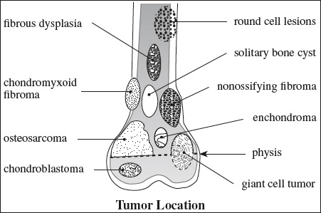
Tumor Position in Transverse Plane
- CENTRAL MEDULLARY LESION
- Enchondroma
- Solitary bone cyst
- ECCENTRIC MEDULLARY LESION
- Giant cell tumor
- Osteogenic sarcoma, chondrosarcoma, fibrosarcoma
- Chondromyxoid fibroma
- CORTICAL LESION
- Nonossifying fibroma
- Osteoid osteoma
- PERIOSTEAL / JUXTACORTICAL LESION
- Juxtacortical chondroma / osteosarcoma
- Osteochondroma
- Parosteal osteogenic sarcoma

Tumor Position in Longitudinal Plane
- EPIPHYSEAL LESION
- Chondroblastoma (prior to closure of growth plate)
- Intraosseous ganglion, subchondral cyst
- Giant cell tumor (originating in metaphysis)
- Clear cell chondrosarcoma
- Fibrous dysplasia
- Abscess
mnemonic: CAGGIE- Chondroblastoma
- Aneurysmal bone cyst
- Giant cell tumor
- Geode
- Infection
- Eosinophilic granuloma [after 40 years of age throw out “CEA” and insert metastases / myeloma]
- METAPHYSEAL LESION
- Nonossifying fibroma (close to growth plate)
- Chondromyxoid fibroma (abutting growth plate)
- Solitary bone cyst
- Osteochondroma
- Brodie abscess
- Osteogenic sarcoma, chondrosarcoma
- DIAPHYSEAL LESION
- Round cell tumor (eg, Ewing sarcoma)
- Nonossifying fibroma
- Solitary bone cyst
- Aneurysmal bone cyst
- Enchondroma
- Osteoblastoma
- Fibrous dysplasia
mnemonic: FEMALE- Fibrous dysplasia
- Eosinophilic granuloma
- Metastasis
- Adamantinoma
- Leukemia, Lymphoma
- Ewing sarcoma

Tumors Localizing to Hematopoietic Marrow
- Metastases
- Plasma cell myeloma
- Ewing sarcoma
- Histiocytic lymphoma
Diffuse Bone Marrow Abnormalities in Childhood
- REPLACED BY TUMOR CELLS
- metastatic disease
- Neuroblastoma (in young child)
- Lymphoma (in older child)
- Rhabdomyosarcoma (in older child)
- primary neoplasm
- Leukemia
- metastatic disease
- REPLACED BY RED CELLS
= red cell hyperplasia = reconversion- severe anemia: sickle cell disease, thalassemia, hereditary spherocytosis
- chronic severe blood loss
- marrow replacement by neoplasia
- treatment with granulocyte-macrophage colony stimulating factor
- REPLACED BY FAT
- Myeloid depletion = aplastic anemia
- REPLACED BY FIBROUS TISSUE
- Myelofibrosis
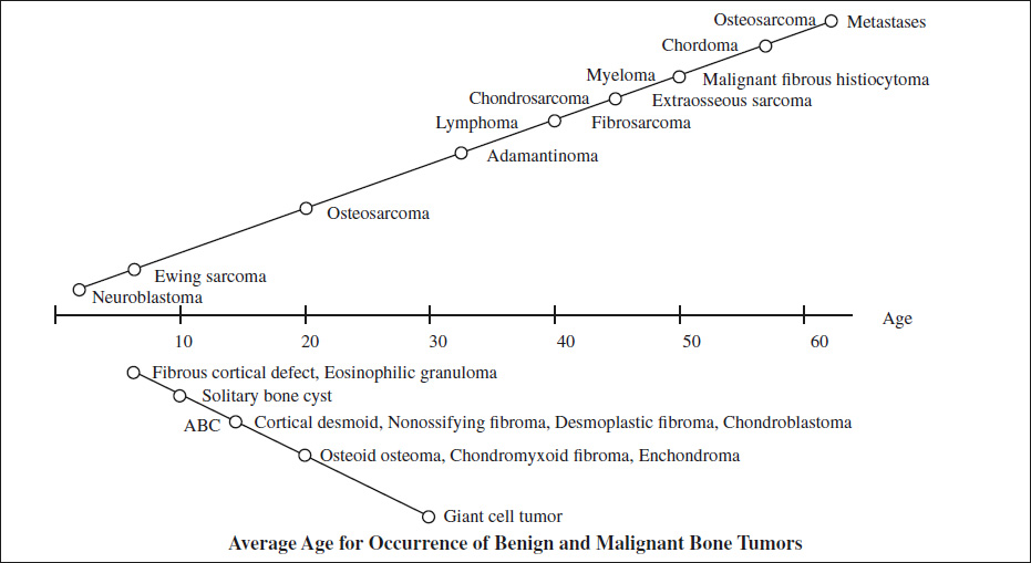
- 80% of bone tumors are correctly determined on the basis of age alone!
Most Frequent Benign Bone Tumor
- Osteochondroma 20–30%
- Enchondroma 10–20%
- Simple bone cyst 10–20%
- Osteoid osteoma
- Nonossifying fibroma
- Aneurysmal bone cyst 5%
- Fibrous dysplasia
- Giant cell tumor
Most Frequent Malignant Bone Tumor
- Bone malignancy
- Metastasis
- Primary bone malignancy
- Multiple myeloma
- Osteosarcoma
- Chondrosarcoma
- Ewing sarcoma
- Primary bone malignancy in children & adolescents
- Osteosarcoma
- Ewing sarcoma
Sarcomas by Age
mnemonic: Every Other Runner Feels Crampy Pain On Moving
- Ewing sarcoma 0 –10 years
- Osteogenic sarcoma 10–30 years
- Reticulum cell sarcoma 20–40 years
- Fibrosarcoma 20–40 years
- Chondrosarcoma 40–50 years
- Parosteal sarcoma 40–50 years
- Osteosarcoma 60–70 years
- Metastases 60–70 years
Ewing Sarcoma Family
- Ewing sarcoma of bone
- Extraskeletal Ewing sarcoma
- Primitive neuroectodermal tumor
- Askin tumor
Malignancy with Soft-tissue Involvement
mnemonic: My Mother Eats Chocolate Fudge Often
- Metastasis
- Myeloma
- Ewing sarcoma
- Chondrosarcoma
- Fibrosarcoma
- Osteosarcoma
Cartilage-forming Bone Tumors
- centrally located ringlike / flocculent / flecklike radiodensity
- BENIGN
- Enchondroma
- Parosteal chondroma
- Chondroblastoma
- Chondromyxoid fibroma
- Osteochondroma
- MALIGNANT
- Chondrosarcoma
- Chondroblastic osteosarcoma
Bone-forming Tumors
- inhomogeneous / homogeneous radiodense collections of variable size + extent
- BENIGN
- Osteoma
- Osteoid osteoma
- Osteoblastoma
- Ossifying fibroma
- MALIGNANT
- Osteogenic sarcoma
Fibrous Connective Tissue Tumors
- BENIGN FIBROUS BONE LESIONS
- cortical
- Benign cortical defect
- Avulsion cortical irregularity
- medullary
- Herniation pit
- Nonossifying fibroma
- Ossifying fibroma
- Congenital generalized fibromatosis
- corticomedullary
- Nonossifying fibroma
- Ossifying fibroma
- Fibrous dysplasia
- Cherubism
- Desmoplastic fibroma
- Fibromyxoma
- Benign fibrous histiocytoma
- cortical
- MALIGNANT
- Fibrosarcoma
Tumors of Histiocytic Origin
- LOCALLY AGGRESSIVE
- Giant cell tumor
- Benign fibrous histiocytoma
- MALIGNANT
- Malignant fibrous histiocytoma
Tumors of Fatty Tissue Origin
- BENIGN
- Intraosseous lipoma
- Parosteal lipoma
- MALIGNANT
- Intraosseous liposarcoma
◊Lipomas follow the SI of subcutaneous fat in all sequences!
Tumors of Vascular Origin
<1% of all bone tumors
- BENIGN
- Hemangioma
- Glomus tumor
- Lymphangioma
- Cystic angiomatosis
- Hemangiopericytoma
- MALIGNANT
- Malignant hemangiopericytoma
- Angiosarcoma = hemangioendothelioma
Metastatic sites: lung, brain, lymph nodes, other bones
Tumors of Neural Origin
- BENIGN
- Solitary neurofibroma
- Neurilemmoma
- MALIGNANT
- Neurogenic sarcoma = malignant schwannoma
Bone Tumors with Fluid-Fluid Levels
- Aneurysmal bone cyst
- Telangiectatic osteosarcoma
- Giant cell tumor
- Chondroblastoma
- Fibrous dysplasia
Round Cell Tumor
Location: arises in midshaft
- osteolytic lesion
- reactive new bone formation
- NO tumor new bone
mnemonic: LEMON
- Leukemia, Lymphoma
- Ewing sarcoma, Eosinophilic granuloma
- Multiple myeloma
- Osteomyelitis
- Neuroblastoma