section name header

Image

Treatment of Deep Venous Thrombosis

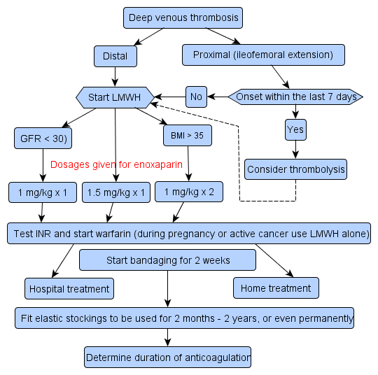
!!flowchart!!

Deep vein thrombosis

Essentials

  • As many as half of all cases of deep vein thrombosis (DVT) develop without clinical symptoms, and only pulmonary embolism (PE) prompts diagnostic tests.
  • A normal D-dimer test result is enough to rule out DVT when, based on clinical presentation, the probability of DVT is no more than moderate. However, if the likelihood of DVT is clinically high, diagnostic imaging studies are indicated.
  • Before treatment is started, a blood sample should be collected for the analysis of blood clotting factors (thrombophilia screening Evaluation of Thrombophilia and Prevention of Thrombosis) if the patient has a positive family history, recurrent or idiopathic (no identified risk factors) thrombosis, massive thrombosis, miscarriage or thrombi affecting both the venous and arterial vessels, or if the patient is young.
  • Treatment aims to prevent PE and post-thrombotic syndrome.
  • All risk factors, or their absence, must be recorded. They determine the duration of the anticoagulant therapy.

Risk factors

  • DVT is rare if no risk factors are present.
  • The most important risk factors for DVT are
    • previous venous thrombosis or embolism
    • severe infection, heart failure
    • oral contraceptives
    • oestrogen therapy or pregnancy
    • immobility (bed rest, flight travel, fractures)
    • surgery
    • cancer
    • inherited thrombophilia Evaluation of Thrombophilia and Prevention of Thrombosis.
  • All risk factors, or their absence, must be recorded. They determine the duration of the anticoagulant therapy (3 months - indefinite).

Clinical assessment

Clinical picture

  • Common signs and symptoms associated with lower limb DVT are:
    • oedema Leg Oedema, pain
    • dilatation of superficial veins
    • Positive Homan's sign (flexion of the ankle causes calf pain).
  • As many as half of all cases of DVT develop without clinical symptoms, and only PE prompts diagnostic tests. However, the specificity of the above signs and symptoms is small, particularly when they occur alone .
  • In addition to the lower limbs, venous thrombosis may also develop occasionally in
    • an upper limb
    • the pelvic veins
    • association with a central venous catheter
    • the right heart chambers
    • the portal vein and cerebral venous sinuses.

Differential diagnosis

  • Alternate diagnoses to be considered in the differential diagnosis include

Assessment of pretest probability

  • The scoring of pretest probability of DVT is presented in table T1.

Assessment of pretest probability

Clinical parameterScore
Active cancer (treatment ongoing, within 6 months or palliative)1
Paralysis, paresis or recent plaster immobilisation of a lower limb1
Recently bedridden for longer than 3 days or major surgery within 4 weeks1
Localised tenderness along the distribution of the deep venous system1
Entire leg swollen1
Calf swelling>3 cm compared with the asymptomatic leg (measured 10 cm below the tibial tuberosity)1
Pitting oedema (greater in the symptomatic leg)1
Collateral superficial veins (non-varicose)1
Alternative diagnosis as likely or greater than that of DVT- 2
  • 3 points or more = high probability, about 75% risk of DVT
  • 1-2 points = moderate probability, about 17% risk of DVT
  • 0 points = low probability, about 3% risk of DVT
Wells PS, Anderson DR, Bormanis J et al. Value of assessment of pretest probability of deep-vein thrombosis in clinical management. Lancet 1997;350:1795-8 1
If the D-dimer test is negative and the score < 3, no other investigations are needed.
If the D-dimer test is positive or the score 3 or higher, compression ultrasonography is indicated.

Investigations

  • It is not always necessary to request imaging studies as an emergency procedure, and they can be carried out during normal work hours.
  • If there is a high suspicion of venous thrombosis, low molecular weight heparin (LMWH) can be started before the investigations.

D-dimer

  • The body's fibrinolytic system is activated in the presence of thrombosis, which results in an increased concentration of D-dimer in the plasma.
  • Elevated D-dimer levels are also present in many conditions other than thrombosis (e.g. severe infection/inflammation, cancer, trauma, surgery, pregnancy). Up to 90% of elderly hospitalised patients have elevated D-dimer concentrations as a consequence of infections and tissue damage.
  • A normal D-dimer test result is enough to rule out DVT when, based on clinical presentation, the probability of DVT is no more than moderate.
  • A normal D-dimer test result together with a negative ultrasonography result does, however, exclude the possibility of DVT with 90-95% probability.

Ultrasonography

  • The investigation is reliable in the diagnosis of proximal DVT (femoral and popliteal veins) but less so in distal DVT.
  • In practice, ultrasonography involving only the femoral and popliteal area is sufficient (2-point ultrasonography, videos Compression Ultrasonography of Veins Deep Venous Thrombosis (Compression Ultrasonography)).
  • If the ultrasonography result is not consistent with the clinical picture and the D-dimer test is positive, ultrasonography should either be repeated in one week's time or venography is indicated.

Venography

  • Venography (a contrast medium examination of the lower limb veins) is indicated if
    • the ultrasonography result is inconclusive
    • laboratory findings are unclear
    • no other explanation can be found for the symptoms.
  • Plasma creatinine must be checked.
  • The radiation exposure is minimal and, for example, pregnancy is not a contraindication to the investigation. However, an obstetrician should be consulted before the investigation is carried out.

Other laboratory tests

  • Before treatment is started, a blood sample should be collected for the analysis of blood clotting factors (thrombophilia screening Evaluation of Thrombophilia and Prevention of Thrombosis) if the following apply to the patient (currently or in the past)
    • a positive family history
    • recurrent or idiopathic (no identified risk factors) thrombosis
    • massive thrombosis
    • a young patient
    • miscarriage
    • thrombi affecting both the venous and arterial vessels.

Treatment

  • The aim of treatment in lower limb DVT is to prevent
    • PE Pulmonary Embolism
    • the development of lower limb vein insufficiency.
  • The decision on whether DVT can be treated in the primary health care depends on the clinical picture and the patient's home situation.
  • Most cases are treated primarily at the patient's home.
  • Hospital treatment is required for
    • DVT with severe symptoms
    • submassive and massive PE.

Management in primary care

  • The treatment of both DVT with only a few symptoms and mild PE can be carried out at a health centre, by a district nurse or self administered by the patient. Based on the individual situation, the treating physician will decide where the treatment should be carried out.
    • Obese patients will need two daily injections because of the large doses needed.
    • Patients with several comorbidities or renal failure are usually not suitable for home treatment.
    • The patient must be supplied with written instructions on how to carry out the treatment.
  • If treatment is carried out at home, the following must be ensured:
    • correct injection technique and correct dose
    • adequate monitoring of the anticoagulant therapy
    • instructions regarding compression bandages and stockings
    • monitoring the patient for possible complications (bleeding, emboli)
  • A follow-up appointment should be made at the latest when the anticoagulant therapy is about to finish.
    • The patient is asked about his/her health and checked for signs of recurrence and post-thrombotic syndrome.

Anticoagulant therapy: dose and duration

  • LMWH is principally used to treat
    • DVT below the knee and at the thigh level
    • more proximal thrombi provided no severe symptoms are present
  • Dalteparin by subcutaneous injection 100 units/kg twice daily or 200 units/kg once daily.
  • Enoxaparin by subcutaneous injection 1 mg/kg twice daily or 1.5 mg/kg once daily.
  • Warfarin is started concomitantly, either 5 mg/day or with the estimated maintenance dose for 3 days and then as guided by INR readings.
  • Heparin is continued
    • until INR has been within the target range (2.0-3.0) for 2 days
    • in any case for at least 5 days.
  • Fondaparinux is an alternative for LMWH. It is suitable for patients with heparin allergy and for the treatment of heparin-induced thrombocytopenia (HIT).
  • LMWH is suitable during pregnancy. Breast feeding is not a contraindication to warfarin.
  • Patients treated for active cancer can be managed with LMWH for 3-6 months followed by warfarin.
  • Duration of anticoagulant therapy, see table T2

Duration of anticoagulant therapy

IndicationsDuration
First episode of thrombosis with a transient risk factor present (e.g. surgery, trauma, immobility, hormonal contraception or replacement therapy, pregnancy)3-6 months
First episode of unprovoked thrombosisAt least 6 months
First episode of thrombosis in a patient with
  • cancer
  • anticardiolipin antibodies or lupus anticoagulant in repeated tests (with a 3 month interval)
  • homozygous factor V or prothrombin (factor II) gene mutation
  • established antithrombin deficiency
  • established protein C or S deficiency
  • combination of two or more thrombophilias
Indefinite
Recurrent unprovoked thrombosisIndefinite

Thrombolytic therapy (fibrinolytic therapy)

  • Thrombolysis may be attempted if the thrombus
    • is recent (less than 1 week) and
    • extends above the inguinal ligament or proximally in an upper limb thrombosis and
    • causes severe symptoms and significant oedema.
  • Thrombolysis can be considered if all the above criteria are fulfilled and the patient is not at an increased risk of bleeding.
  • Local, catheter-directed thrombolysis is the treatment of choice if a radiologist competent in the procedure is available.

Surgical treatment

  • Surgery is the first-line treatment approach if the viability of the limb is threatened and particularly if both thrombolytic and anticoagulant therapy are contraindicated.

Other treatment

  • Immediate bandaging during the acute phase to ensure the competence of the communicating veins
    • Using an elastic bandage, the leg is bandaged from the foot to the knee gradually decreasing the pressure as the dressing advances proximally.
    • The bandage is applied in circular turns; a figure of eight bandage is too tight.
    • Catheter-directed thrombolysis is not a contraindication to bandaging.
    • If the swelling extends to the thigh, the leg should be bandaged up to the groin.
  • The patient should mobilise as soon as clinically possible.
  • Follow-up treatment with graduated compression stockings (Class II) for at least 2 years reduces the likelihood of post-thrombotic syndrome .
  • Patient education

Related resources

  • Cochrane reviews
  • Other evidence summaries
  • Clinical guidelines
  • Literature