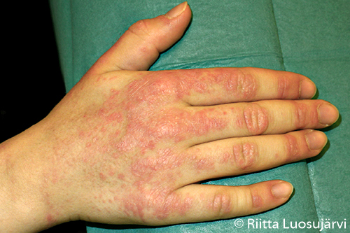
Information

Editors

RistoNikku
MarttiTeikari

Dislocation of the Patella

Mechanism of injury

  • Valgus bend and external rotation of the lower leg while the knee is flexed causes the patella to dislocate over the lateral crest of femoral sulcus.

Symptoms and findings

  • A typical patient is a teenager or a young adult with haemarthrosis of the knee (at first dislocation; habitual dislocations are not associated with haemarthrosis).
  • History (slipping of the patella noticed by the patient him-/herself: the energy leading to the dislocation may vary; family history)
  • Difficulty in weight bearing
  • Pain and palpable tenderness on the medial side of the patella, on the medial epicondyle of the femur and on the upper crest of the lateral condyle of the femur. Sometimes the dislocation is visible.
  • The patella can be displaced laterally more on the injured side compared with the intact side. This procedure is painful (the apprehension sign).
  • X-ray (including the patellar axial projection) is necessary to find any bony fragments. Patellar axial projection may reveal tilting and lateralization of the patella (pictures Determining the Position of the Patella Patellar Dislocation).
    • In profuse hemarthrosis of the knee, MRI should be considered..

Treatment Surgical Vs. Non-Surgical Interventions for Patellar Dislocation

Reduction

  • The knee is extended and the patella is pressed in medial direction.

Evacuation of the haemarthrosis

  • Major haemarthrosis is aspirated, which alleviates the pain. Presence of fat droplets suggests osteochondral fracture.

Indications for operative treatment

  • Loose body on x-ray needing fixation
  • Operative treatment of recurrent dislocations is considered depending on the age of the patient and the frequency of dislocations. The key question is: Do you trust your knee?
  • The extent of patellar articular cartilage lesions and prolongation of follow-up time appear to correlate with the treatment results.

Conservative treatment

  • Primary dislocation with only mild haemarthrosis
  • Acute phase of recurrent dislocation. An operation is performed later if necessary.
  • Quadriceps exercises are started and a patellar stabilizing orthosis is used when the oedema has diminished (in 2-3 days).
  • Short immobilization may be used to relieve pain.

    References

    • Pidoriano AJ, Weinstein RN, Buuck DA, Fulkerson JP. Correlation of patellar articular lesions with results from anteromedial tibial tubercle transfer. Am J Sports Med 1997 Jul-Aug;25(4):533-7. [PubMed]
    • Christiansen SE, Jakobsen BW, Lund B, Lind M. Isolated repair of the medial patellofemoral ligament in primary dislocation of the patella: a prospective randomized study. Arthroscopy 2008 Aug;24(8):881-7. [PubMed]
    • Palmu S, Kallio PE, Donell ST, Helenius I, Nietosvaara Y. Acute patellar dislocation in children and adolescents: a randomized clinical trial. J Bone Joint Surg Am 2008 Mar;90(3):463-70. [PubMed]
    • Sillanpää PJ, Mattila VM, Mäenpää H, Kiuru M, Visuri T, Pihlajamäki H. Treatment with and without initial stabilizing surgery for primary traumatic patellar dislocation. A prospective randomized study. J Bone Joint Surg Am 2009 Feb;91(2):263-73. [PubMed]
    • Camanho GL, Viegas Ade C, Bitar AC, Demange MK, Hernandez AJ. Conservative versus surgical treatment for repair of the medial patellofemoral ligament in acute dislocations of the patella. Arthroscopy 2009 Jun;25(6):620-5. [PubMed]
    • Nikku R, Nietosvaara Y, Aalto K, Kallio PE. The mechanism of primary patellar dislocation. Acta Orthop 2009 Jan 1;80(4):1-3. [PubMed],
    • Yao LW, Zhang C, Liu Y et al. Comparison operative and conservative management for primary patellar dislocation: an up-to-date meta-analysis. Eur J Orthop Surg Traumatol 2015;25(4):783-8. [PubMed]

Related Resources

Related resources