Central Retinal Artery Occlusion
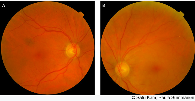
Central retinal artery occlusion. The right eye (Figure A) is normal except for arteriolosclerotic changes. The left eye (picture B) has a sudden loss of vision. Central retinal artery thrombosis has caused milky white ischaemic swelling of the retina in the area between the blood vessel arches on the temporal side. A cherry red spot is visible in the foveola in the centre of the macula. The embolus has migrated into the superior temporal arterial branch and stopped at the junction, but the migration has occurred too late to save the patient's central vision.
Picture and text: Satu Kam and Paula Summanen. [Central retinal artery occlusions]. In: Seppänen M, Kaarniranta K, Setälä N, Uusitalo H (eds.). [Handbook of ophthalmology]. Helsinki: Duodecim Publishing Company 2022.
Primary/Secondary Keywords
- retina
- central retinal artery occlusion
- central artery occlusion
- CRAO
- eye
- macula
- macula retinae
- vision
- vision loss
- sudden vision loss
- loss of vision
- sudden loss of vision