Lobar Pneumonia (Lateral View)
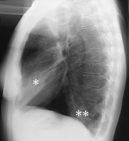
Lobar pneumonia (lateral view). Lobar pneumonia. The lateral radiograph shows alveolar consolidation both in the middle lobe (*) and in the inferior lobe (**) of the right lung. For PA view: see Lobar Pneumonia (Pa View).
Picture: Duodecim Medical Publications Ltd
Primary/Secondary Keywords