Trochanteric Hip Fracture

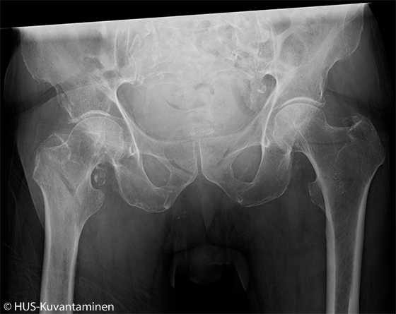
Trochanteric hip fracture (PA radiograph without markers).
An old man fell at home and injured his right hip. Physical examination revealed a shortening of the right lower limb, and the limb was in external rotation.

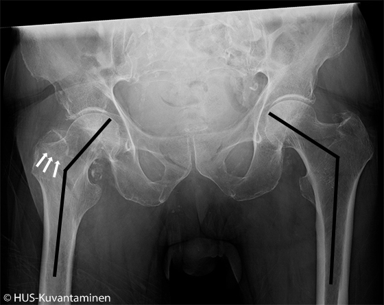
Trochanteric hip fracture (PA radiograph with markers).
In plain x-ray of the pelvis, the angle between the head and diaphysis of the femur is reduced on the right side and there is also a pretrochanteric sclerotic line of impaction visible.
Picture and text: Tiina Lehtimäki, HUS Imaging

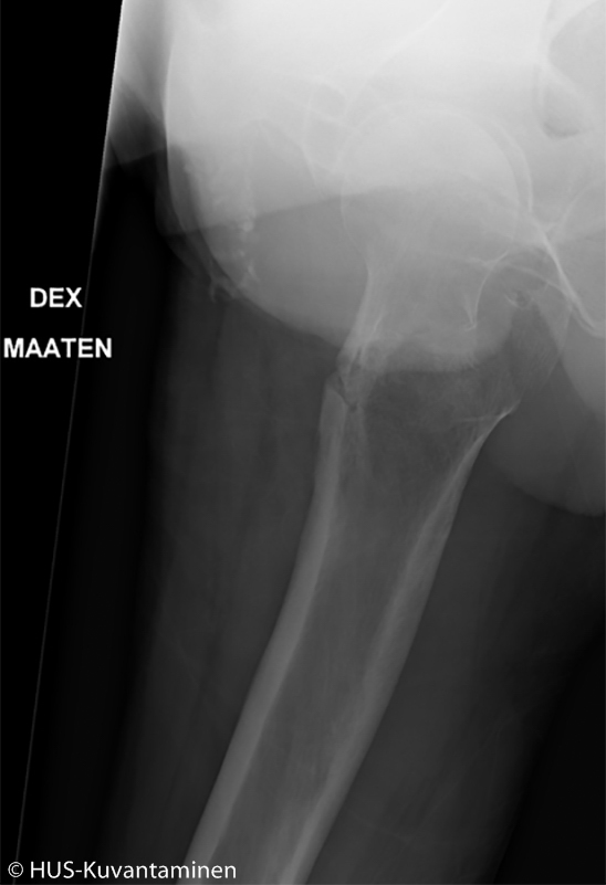
Trochanteric hip fracture (lateral radiograph without markers).
Picture and text: Tiina Lehtimäki, HUS Imaging

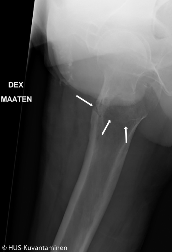
Trochanteric hip fracture (lateral radiograph with markers).
The lateral radiograph shows comminuted bone cortex and a radiolucent line caused by the fracture.
Picture and text: Tiina Lehtimäki, HUS Imaging
Primary/Secondary Keywords
- hip
- fracture
- trochanteric fracture
- pelvis
- pelvis x-ray
- x-ray