Intense Cryotherapy of Superficial Basal Cell Carcinoma on the Back - Image Series
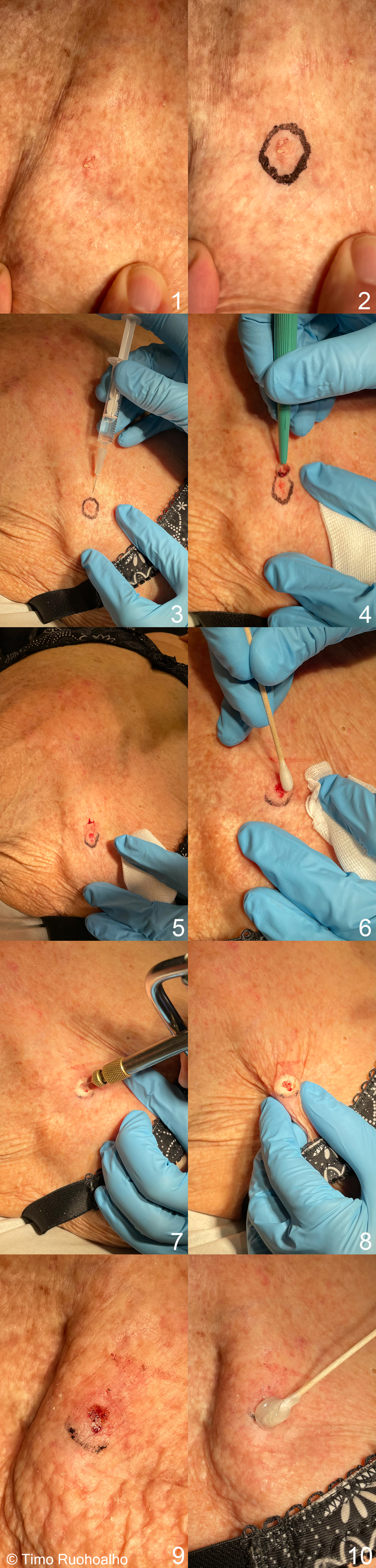
Intense cryotherapy of superficial basal cell carcinoma on the back - image series. Diagnose the lesion (in this case carcinoma in situ) and use a marker to draw the outline of the lesion to be treated and the freeze margin. Usually, about 3 mm freeze margins are used (Images 1 and 2).
After marking the area, disinfect the area to be treated and infiltrate a local anaesthetic (Image 3).
When the area is numb, carefully remove any abnormal tissue with a curette (Images 4 and 5).
Oozing of blood can be stopped by pressing gently with a cotton swab dipped in ferric chloride or aluminium chloride solution.
Freeze the lesion white to establish a halo thaw time (to the border of the lesion) of one minute. Allow the entire lesion to thaw properly, and then repeat freezing (Images 7 and 8).
There will be a weeping superficial ulcer and a clear blister (Image 9). It is advisable to apply, for example, fucidic acid ointment to the area daily for 1-2 weeks (Image 10).
The treatment practically always leaves a permanent light scar that will be quite visible on dark skin. Intense cryotherapy technique can be used by a medical specialist or a physician with special expertise in cryotherapy, as necessary.It must never be used below knee level, as this can easily lead to chronic ulceration in that area. It should basically not be used in primary health care for lesions in the head or neck area at all.When treating lesions in the head area in specialized care, the cosmetic and functional results and risk of recurrence should be carefully considered. The treatment result should be assessed by a check-up visit after about 3-6 months.
See also video Intense Cryotherapy of Superficial Basal Cell Carcinoma on the Back - Video.
Pictures: Timo Ruohoalho, text: Alexander Salava and Timo Ruohoalho
Primary/Secondary Keywords
- cryotherapy
- basal cell carcinoma
- intense cryotherapy