Naevus Excision
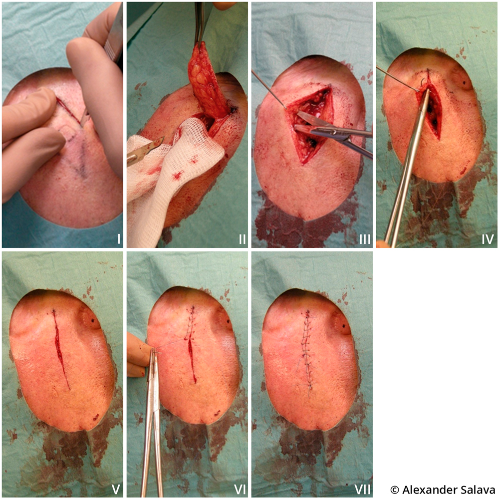
Naevus excision.Naevi should be excised using a fusiform (elliptical) incision with pre-planned clinical margins (image I). The clinical margin is the distance between the lateral edge of the skin lesion and the surgical incision (usually in millimetres). After removal, the sample shrinks and hence the histological margin reported by the pathologist is always smaller than the clinical surgical margin. It is important to remember to cut in a 90-degree angle, ensuring that the naevus is removed all the way to subcutaneous fat (image II). Use scissors to release the wound edges bluntly for a sufficiently long distance to make sure that the wound can be closed with only moderate tension (image III). The ends of the wound area should also be released to reduce the formation of dog ears. For good results an absorbable intracutaneous suture should be used (image IV). This is recommended whenever the area is under high tension. The absorbable intracutaneous suture ensures that the wound stays closed after removal of the skin suture, and facilitates early removal of the superficial suture (image V). In the procedure shown here, a continuous locked suture was used for superficial closure (images VI and VII).
Pictures and text: Alexander Salava
Primary/Secondary Keywords
- suture techniques
- naevus excision
- continuous locked suture
- wound closure
- wound
- wound treatment