Treatment of Cerebral Infarction (a Patient Case)
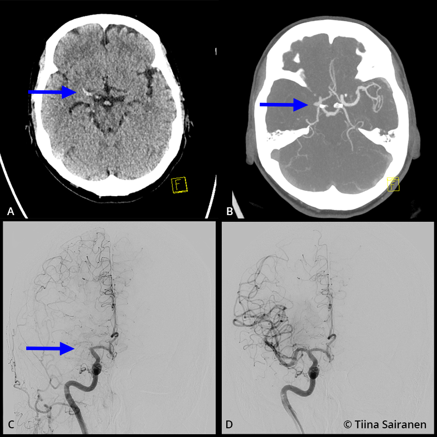
Treatment of cerebral infarction (a patient case). Patient history: A 61-year-old woman living with her spouse, still working. Four years ago curatively treated breast cancer with resection, radiotherapy and hormone therapy. Also hypertension and arrhythmias, smoker, drinks wine at weekends. Last feeling of arrhythmia a week earlier. Current symptom: At work at 13.50 after coughing, left sensorimotor hemiparesis and headache. On recanalisation assessment, NIHSS score of 13.
Imaging shows a dense proximal base of the right middle cerebral artery (MCA), the M1 segment (image A, arrow), subtle loss of grey-white matter differentiation in the right basal ganglia area and widespread sulcal effacement is seen in the right middle cerebral artery vascular territory. No clearly demarcated fresh infarction is seen. On CT angiography, the right MCA terminates bluntly at the base in line with the thrombus (image B, arrow). The patient received thrombolytic therapy at 14.57 (1 h 7 min from symptom onset) after which thrombectomy was performed with complete opening of the occlusion (image C, arrow) obtained by first aspiration at 15.35 (image D). After the procedure the patient became asymptomatic, NIHSS score 0.
Additional note: NIHSS is a score used to assess cerebral infarction, with values between 0 (normal) and 42. The National Institutes of Health Stroke Scale (NIHSS) score refers to the severity of symptoms measured by a 13-item assessment. A score of 0 = no symptoms, 1-4 = mild symptoms, 5-15 = moderate symptoms, 16-20 = moderate to severe symptoms, 21-42 = severe symptoms.
Picture and text: Tiina Sairanen
Primary/Secondary Keywords
- Cerebral infarction
- Stroke
- ischaemic stroke
- NIHSS
- NIHSS score
- NIHSS scoring
- Thrombolysis
- Thrombectomy
- Hypertension
- Arrhythmias
- Arrhythmia
- Imaging
- CT angiography
- Sensorimotor hemiparesis