Trochanteric Hip Fracture

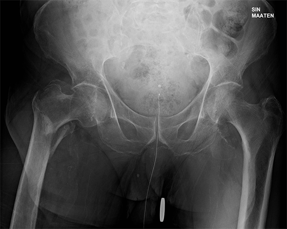
Trochanteric hip fracture (PA radiograph without markers).
A retired man fell in the garden and injured his right lower limb.

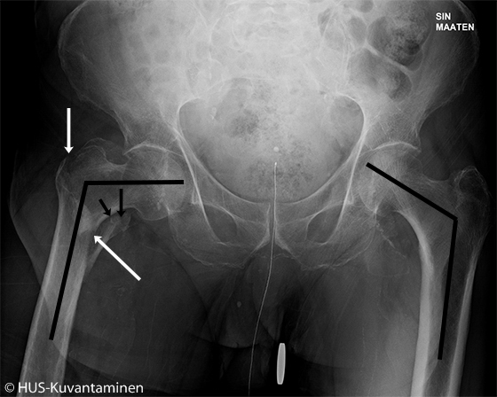
Trochanteric hip fracture (PA radiograph with markers)
A plain x-ray of the pelvis reveals a trochanteric fracture on the right side. Due to inferior displacement of the femoral head, the angle between the femoral head and diaphysis is reduced (varus malposition). On both sides of the trochanter, bone cortex is discontinuous (black arrows). The bone structure shows increased translucency and looks osteoporotic.
Picture and text: Tiina Lehtimäki, HUS Imaging
Primary/Secondary Keywords
- hip
- femur
- femur fracture
- hip fracture
- trochanter
- osteoporosis
- hip x-ray
- pelvis x-ray