Becker's Naevus
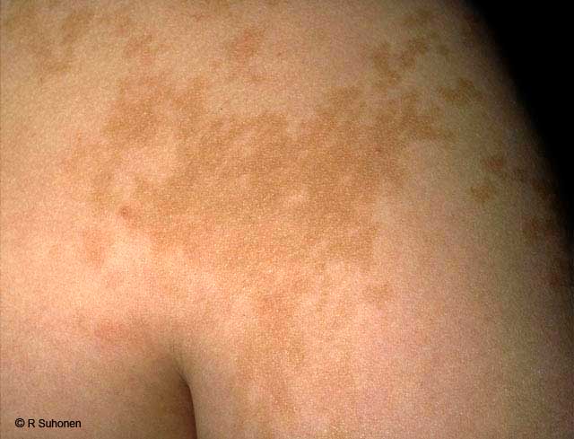
Becker's naevus. Becker's naevus (melanosis naeviformis Becker-Siemens) resembles pityriasis versicolor. The patch does not scale at all (when scraped), the periphery is "archipelago-like", and in boys the patch usually has a more pronounced hair growth than usual. See also pictures Becker's Naevus with Increased Hair Growth and Becker's Melanosis on the Back.
Picture and text: Raimo Suhonen
Primary/Secondary Keywords
- dermatology
- Becker's naevus
- melanosis naeviformis Becker-Siemens
- Becker's melanosis
- Becker naevus
- Becker nevus
- shoulder