Patellar Dislocation
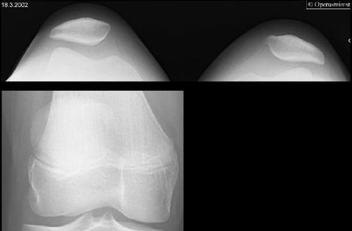
Patellar dislocation. The knee of a 12-year-old girl was twisted while skiing. In clinical examination there was considerable tenderness on the medial side of the left knee as well as significant soft tissue swelling. The radiographs show the patellar projections of both knees and plain x-ray of the left knee: the left patella is clearly lateralized. The tibiofemoral space is normal, all the bony joint surfaces are intact and no fractures are seen. Treatment consisted of a Camp patellar support and instruction by a physiotherapist. A follow-up appointment after 1 month was agreed on. In suspected luxation the radiological investigations include anterior and lateral projections of the affected knee as well as axial projection of both knees in 20-30° flexion (so-called patellar or Laurin's projection). The patellar projection reveals the lateral displacement and tilting of the patella, suggesting luxation. Avulsion fractures of the medial margin of the patella, as well as joint surface fractures and loose bodies can be diagnosed. However, loose bodies often consist mainly of cartilage containing very little bone and are thus not necessarily visible on x-ray.
Picture: Medimage / University of Turku. By courtesy of the Finnish Ministry of Education.
Primary/Secondary Keywords
- Radiology
- Traumatology
- Patellar dislocation
- Patella
- Patellar luxation
- Patellar projection
- Laurin's projection