section name header

Image

Cryotherapy of Actinic Keratosis

Cryotherapy of actinic keratosis

Define the areas to be treated (Image 1).

Curette thickened actinic keratoses, as necessary (Images 2 and 3). If the skin underneath the hyperkeratosis is clearly thickened or there is raw ulceration, a biopsy sample should be taken.

Oozing of blood can be stopped by applying aluminium or ferric chloride (Image 4).

The lesion should be completely frozen (Image 5) and allowed to stay frozen for a short while. The lesion should remain frozen for 5-20 seconds depending on the area of skin involved (on the face, 5-10 seconds is usually enough); 2-3-mm margin (freeze margin beyond the margin of the skin lesion); usually 2 freeze-thaw cycles per session.

Even after mild cryotherapy, the treated area will be erythematous (Image 6).

A physician with appropriate training can also treat actinic keratoses in primary care. Particularly when treating a patient's first actinic keratoses, the diagnosis should be confirmed histologically (biopsy). It is important to confirm the treatment result by a checkup visit after about 3-6 months.

See also video

Pictures: Timo Ruohoalho, text: Alexander Salava

Primary/Secondary Keywords
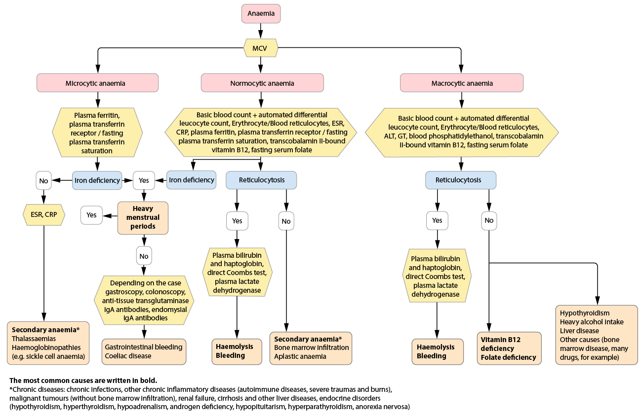