Types of Supraventricular Tachycardia Seen on ECG

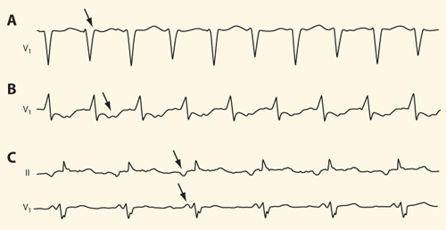
Types of supraventricular tachycardia seen on ECG.A. Atrioventricular nodal re-entry tachycardia (AVNRT). The pathogenic mechanism behind the arrhythmia is a re-entry circuit within the atrioventricular node. The QRS complex is narrow and the ventricular rate is regular 185/min. In typical AVNRT the impulse travels over the so-called slow pathway from the atrium to the ventricle and returns over the so-called fast pathway; this explains the retrograde P wave seen in the lead V1 assimilated to the end of the QRS complex (arrow). The arrhythmia was corrected by severing the slow pathway through catheter ablation.
B. Orthodromic tachycardia (AVRT). The ventricular rate is 152/min. During the arrhythmia, the impulse travels over the normal pathway from the atrium to the ventricle and returns over an accessory conducting pathway, resulting in a narrow QRS complex. The retrograde P wave comes later than in AVNRT and is best seen in the lead V1. The arrhythmia was corrected by severing the accessory conducting pathway through elective catheter ablation.
C. Atrial tachycardia. Ectopic atrial tachycardia with a ventricular rate of 105/min. The P wave is positive in the lead V1 but negative in the lead II, thus excluding sinus tachycardia. In the electrophysiological investigation the ectopic focus was localized to the right atrium just inside the coronary sinus orifice and was successfully treated through catheter ablation.
Picture: Duodecim Medical Publications Ltd
Primary/Secondary Keywords
- Electrocardiography
- Tachycardia
- Re-entry tachycardia
- Orthodromic tachycardia
- Atrial tachycardia
- AVNRT
- AVRT
- Ventricular rate
- Catheter ablation
- Retrograde P wave