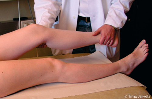
Examining Lateral Stability of the Knee

Examining lateral stability of the knee. Lateral stability should be tested on both the medial and lateral sides, each time with the knee extended and slightly flexed (20-30 degrees). If the completely extended knee gives way, you should suspect injury not only to the collateral ligaments but also to the posterior capsule or cruciate ligaments. If the collateral ligament alone is damaged, the knee will give way only when slightly flexed.
Picture and text: Timo Järvelä
Primary/Secondary Keywords
- knee
- knee injury
- ACL
- PCL
- posterior capsule
- posterior knee capsule
- ACL injury
- PCL injury