Onychomycosis (Tinea Unguium)
Figure 1
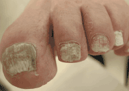
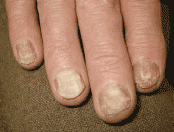
Figure 2
Etiology: Dermatophyte infection of nail plate, often associated with tinea pedis. Less commonly due to yeasts and nondermatophyte molds.
History: Change in nail color and more brittle; usually asymptomatic.
Physical: Yellow discoloration, thickening, nail dystrophy, subungual hyperkeratosis, onycholysis. Toenails > fingernails.
Patterns include the distal subungual form (most common), the proximal white subungual form (may be a sign of HIV disease), and the white superficial form.
Investigations: 20% KOH direct microscopy, nail clipping/scraping for culture.
DDx: Nail psoriasis or trauma, eczema, lichen planus.
- Topical antifungals much less effective. Ciclopirox (Penlac®) nail lacquer may be tried if po Tx not an option.
- Important to culture fungus prior to initiating oral Tx
- Terbinafine (Lamisil) 250 mg po qd × 6 wk for fingernails, × 12 wk for toenails; itraconazole (Sporanox) 200 mg po bid × 7 d, then 3 wk off—2 pulses for fingernails, 3 pulses for toenails.
- Other less common options: Griseofulvin (esp. in kids), fluconazole.
- Hepatotoxicity and leukopenia are rare side effects of terbinafine use, some check baseline and monitor CBC and LFTs during Tx; itraconazole—mostly hepatotoxic S/E.
- Educate patients that due to slow rate of new nail formation, it may take many months after treatment to see clinical improvement.