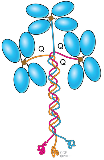
Structure of Acetylcholinesterase

(From Naguib M, Flood P, McArdle JJ, et al. Advances in neurobiology of the neuromuscular junction: implications for the anesthesiologist. Anesthesiology. 2002;96:202-231.)
After an action potential and Ca2+ influx, phosphorylation of synapsin is activated by calcium-calmodulin activated protein kinases I and II. This results in the mobilization of synaptic vesicles (SVs) from the cytomatrix toward the plasma membrane. The formation of the SNARE complex is an essential step for the docking process. After fusion of SVs with the presynaptic plasma membrane, acetylcholine (ACh) is released into the synaptic cleft. Some of the released acetylcholine molecules bind to the nicotinic acetylcholine receptors (nAChRs) on the postsynaptic membrane while the rest is rapidly hydrolyzed by the acetylcholinesterase (AChE) present in the synaptic cleft to choline and acetate. Choline is recycled into the terminal by a high-affinity uptake system, making it available for the resynthesis of acetylcholine. Exocytosis is followed by endocytosis in a process dependent on the formation of a clathrin coat and of action of dynamin. After recovering of SV membrane, the coated vesicle uncoats and another cycle starts again. See text for details. Acetyl CoA, acetylcoenzyme A; CAT, choline acetyltransferase; PK, protein kinase.