section name header

Introduction

To access the stomach, duodenum, or jejunum, a practitioner may place a tube through a patient’s abdominal wall when enteral feedings are needed.1 This procedure may be done surgically or percutaneously.

A gastrostomy or jejunostomy tube is usually inserted during intra-abdominal surgery. The tube may be used for feeding during the immediate postoperative period, or it may provide long-term enteral access, depending on the type of surgery. Typically, the practitioner sutures the tube in place to prevent gastric contents from leaking.

In contrast, a percutaneous endoscopic gastrostomy (PEG) or percutaneous endoscopic jejunostomy (PEJ) tube can be inserted endoscopically without the need for laparotomy or general anesthesia. Typically, the insertion is done in the endoscopy suite or in an interventional radiology department.2 Ultrasound can be used to confirm placement. A PEG or PEJ tube may be used for nutrition, drainage, and decompression. Relative contraindications to endoscopic placement include obstruction (such as an esophageal stricture or duodenal blockage), previous gastric surgery, esophageal or gastric varices, morbid obesity, and ascites.3 These conditions necessitate surgical placement. Long-term feeding devices should be considered when the need for enteral feeding is at least 4 weeks in adults, children, or full-term infants.4

With PEJ tube placement, feedings may begin after 24 hours (or when peristalsis resumes). A PEG tube can be used for feedings within several hours of placement. Current research supports PEG tube usage beginning 4 hours or less after placement in both children and adults.4 Commercially prepared enteral administration sets and pumps allow for continuous formula administration.4

Contraindications to enteral feeding include bowel obstruction, severe ileus, severe upper gastrointestinal bleeding, intractable vomiting or diarrhea, severe hemodynamic instability, gastrointestinal ischemia, and a high output fistula.5 Nursing care for patients receiving intermittent or continuous enteral tube feedings includes providing skin care at the tube site, maintaining the feeding tube, administering the enteral formula, monitoring the patient’s response to feedings, adjusting the feeding schedule, and preparing the patient for self-care after discharge.

Equipment

Equipment

For Intermittent or Continuous Feeding

Fluid-impermeable pad or towel • enteral syringe (20-mL or larger)4 • prescribed enteral formula • tap or purified (sterile, distilled, ultrafiltrated, or ultraviolet-light treated) water4 • enteral administration set • IV pole • gloves • labels that state ENTERAL USE ONLY-NOT FOR IV USE • oral care supplies • tape • stethoscope • disinfectant pad • cleaning supplies for syringe • Optional: pH testing equipment, enteral feeding pump, enteral feeding bag, scale, facility-approved disinfectant, tape measure, graduated container.

For Site Care

4" × 4" (10 cm × 10 cm) gauze pads • soap and water or normal saline solution • water • cotton-tipped applicators • hypoallergenic tape • gloves • label • Optional: external stabilization device, sterile gauze or foam dressing, skin protectant, tape measure.

Preparation of Equipment

Preparation of equipment

Allow the enteral formula to warm to room temperature before administration, because feeding the patient room-temperature formula may reduce the risk of diarrhea.6,7

Inspect all equipment and supplies. If a product is expired, is defective, or has compromised integrity, remove it from patient use, label it as expired or defective, and report the expiration or defect as directed by your facility.

Implementation

Implementation
  • Verify the practitioner’s order, including the patient’s identifiers, the prescribed route based on the enteral tube’s tip location, enteral feeding device, prescribed enteral formula, administration method, volume and rate of administration, and type, volume, and frequency of water flushes.4
  • Review the patient’s medical record to make sure that catheter placement was confirmed before beginning the feeding.4
  • Gather and prepare the necessary equipment and supplies.
  • Visually inspect the enteral formula for damage to the container, altered formula characteristics, and expiration date. Don’t use the formula if its integrity is compromised or if it’s expired; instead, obtain a new container of the formula.4
  • Compare the label on the enteral formula container to the order in the patient’s medical record.4
  • Perform hand hygiene.8,9,10,11,12,13
  • Confirm the patient’s identity using at least two patient identifiers.14
  • Provide privacy.15,16,17,18
  • Explain the procedure to the patient and family (if appropriate) according to their individual communication and learning needs to increase their understanding, allay their fears, and enhance cooperation.19
  • Raise the patient’s bed to waist level before providing care to prevent caregiver back strain.20
  • Assess the patient to determine the risk for aspiration.4,21
  • Position the patient with the head of the bed elevated to at least 30 degrees or upright in a chair to prevent aspiration. If this position is contraindicated, consider a reverse Trendelenburg position.4,22
  • Perform hand hygiene.8,9,10,11,12,13
  • Put on gloves to comply with standard precautions.23,24,25

For an Intermittent or a Continuous Feeding

  • Open an enteral feeding bag with enteral administration set tubing if using an open administration system. If using a closed system, open the enteral administration set tubing.
  • Pour only a premeasured 4-hour volume of enteral formula into the enteral feeding bag and hang the bag on the IV pole.4 Set infusion time for open enteral nutrition feeding systems to 4 to 8 hours.4 Limiting the volume decreases the risk of bacterial overgrowth in the formula. Alternatively, open the sterile enteral formula container and attach it to the enteral administration set.
  • If using an enteral feeding pump, attach the enteral administration set tubing to the enteral feeding pump following the manufacturer’s instructions.
  • Prime the enteral administration set according to the manufacturer’s recommendations to minimize air delivery into the GI tract.
  • Make sure that the enteral formula container is labeled with the patient’s identifiers; formula name (and strength if diluted); date and time of formula preparation; date and time that the formula was hung; administration route, rate, and duration (if cycled or intermittent); initials of who prepared, hung, and checked the enteral formula against the order; expiration date and time; dosing weight (if appropriate); and notation ENTERAL USE ONLY-NOT FOR IV USE.4
  • Label the enteral administration set with the date and time that it was first hung. Change the open-system administration set according to the manufacturer’s instructions to prevent bacterial growth. If you’re using a closed system, change the administration set according to the manufacturer’s instructions.4
  • Place a fluid-impermeable pad or towel under the patient’s feeding tube to prevent soiling of linens.
  • Verify tube placement by using at least two of the following methods.4,22,26 Observe for a change in the external length or incremental marking on the tube at the exit site to determine whether the tube has migrated.4,22,26 Observe for a change in volume of aspirate from the feeding tube, because a large increase in volume may signal the upward dislocation of a small-bowel feeding tube into the stomach; persistent inability to withdrawal fluid (or only a few drops of fluid) from the tube may signal upward displacement of a gastric tube into the esophagus.22,26 Review routine chest and abdominal X-ray reports.4,22,26 Aspirate tube contents and inspect the visual characteristics of the tube aspirate, because fasting gastric secretions often appear grassy-green or brown or clear and colorless.4,26 If performed at your facility, measure the pH of aspirate from the tube; fasting gastric pH is usually 5 or less, even in patients receiving gastric acid inhibitors.4,22,26
  • If you suspect tube migration, don’t administer the enteral feeding. Instead, notify the practitioner.
  • Flush the enteral feeding tube with at least 30 mL of water, as ordered. Use purified water for immunocompromised or critically ill patients.4
  • Connect the end of the enteral administration set tubing to the distal end of the enteral tube. Trace the tubing from the patient to its point of origin to ensure that it’s connected to the proper port before beginning the tube feeding.4,27,28
  • Tape the connection to prevent accidental disconnection of the tubing. Route the enteral administration set tubing toward the patient’s feet and place the enteral feeding pump toward the foot of the bed, because a standardized approach to keeping IV lines routed toward the head and enteric lines routed toward the feet prevents dangerous misconnections.27,28 If the patient has different access sites or several bags hanging, label each tubing at the distal end (near the patient connection) and proximal end (near the source container) to distinguish different tubing and prevent dangerous misconnections.28
  • Open the administration set clamp. Regulate the flow to the desired rate. If using an enteral feeding pump, follow the manufacturer’s instructions for setting the flow rate and starting the infusion.4 Make sure that the enteral feeding pump alarm limits are set according to the patient’s current condition, and that alarms are turned on, functioning properly, and audible to staff.29,30,31
  • Monitor the gravity drip rate or enteral feeding pump infusion rate frequently to ensure accurate delivery of the enteral formula.
  • Flush the enteral feeding tube every 4 hours with at least 30 mL of water (purified water for immunocompromised or critically ill patients), as ordered and tolerated, to maintain patency and provide hydration.4
  • Monitor the patient at least every 4 hours for appropriate positioning.4
  • Assess for GI intolerance of enteral tube feedings every 4 hours by assessing for abdominal distention, monitoring for reports of abdominal pain, and observing for passage of flatus and stool.4,22 Gastric residual volume (GRV) may not need to be used routinely to monitor critically ill patients receiving enteral nutrition.4,21 For patient care areas that still monitor GRV, avoid holding the enteral feeding for a GRV of less than 500 mL if the patient has no other signs of feeding intolerance to prevent inappropriate stoppage of enteral feedings.4,32
  • Perform oral care routinely to decrease oral bacterial colonization and, subsequently, reduce the risk of health care–acquired pneumonia.33 (See the "Oral care" procedure.)
  • Remove and discard the fluid-impermeable pad or towel.25
  • Monitor the patient’s weight and nutritional, fluid, electrolyte, and metabolic statuses, as ordered, to evaluate the effectiveness of enteral feedings.

For Site Care

  • Perform hand hygiene.8,9,10,11,12,13
  • Put on gloves to comply with standard precautions.23,24,25
  • Gently remove the dressing to prevent skin stripping or tearing and discard it in an appropriate receptacle.19,34 Don’t cut away the dressing over the catheter, because you might cut the tube or the sutures holding the tube in place.
  • Remove and discard your gloves.23,24
  • Perform hand hygiene.8,9,10,11,12,13
  • Put on a new pair of gloves.23,24

Gastrostomy or Jejunostomy Tube Site

  • Assess the tube exit site for new or increasing pain and signs of skin breakdown, redness, edema, induration, and bleeding.4
  • Inspect the tube for wear and tear. A tube that has worn out needs to be replaced.3,4
  • Observe for a change in external tube length or the incremental marking at the exit site to assess for tube migration.3,4
  • Until healing occurs, clean the skin immediately around the gastrostomy or jejunostomy tube’s exit site daily (and as needed) using a cotton-tipped applicator moistened with normal saline solution. Next, using a 4" × 4" (10 cm × 10 cm) gauze pad soaked in normal saline solution, clean the adjacent skin and pat it dry using another gauze pad. When the exit site has healed, wash the skin around it with soap and water daily. Rinse the area with water and pat it dry.3
  • Apply skin protectant, if necessary, to prevent skin maceration.3
  • Secure the gastrostomy or jejunostomy tube to the skin with and external stabilization device or hypoallergenic tape to prevent peristaltic migration of the tube and tension on the suture that anchors the tube in place.35
  • Coil the tube, if necessary, and tape it to the abdomen to prevent pulling and contamination of the tube. Rotate the taping site to prevent skin damage.

PEG or PEJ Site Care

  • Slide the tube’s outer bumper carefully away from the skin about ½" (1.3 cm). Depress the skin surrounding the tube gently and inspect for leakage. Minimal wound drainage, which appears initially after implantation, should subside in about 1 week.
  • Assess the skin at the exit site for increasing pain and signs of infection, such as redness, edema, and purulent drainage.3
  • Inspect the tube for wear and tear. A tube that is worn out needs to be replaced.3,4
  • Observe for a change in external tube length or the incremental marking on the tube at the exit site to assess for tube migration.3,4
  • Clean the exit site with soap and water-moistened gauze pads (as shown below), and allow it to dry.3,36,37

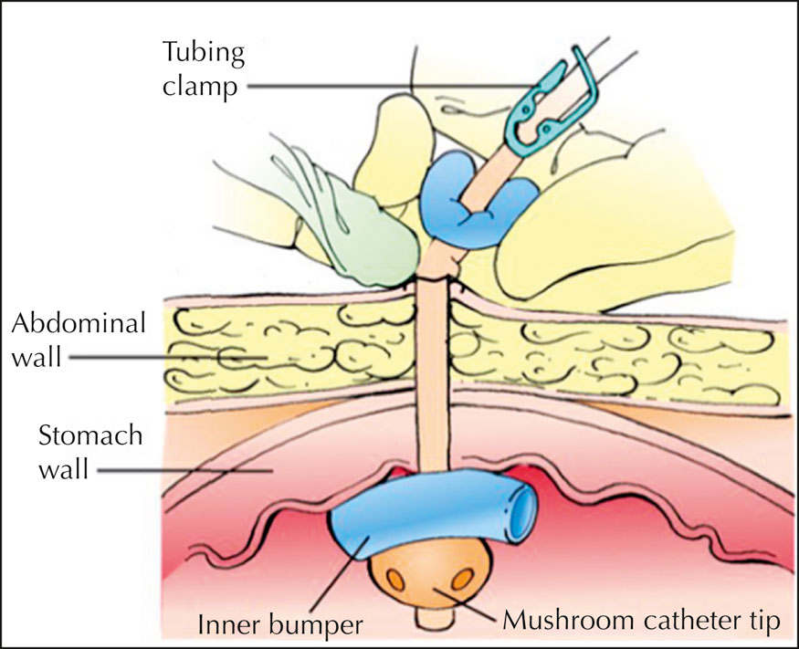
  • Rotate the outer bumper 90 degrees to avoid applying the same tension to the same skin area and to prevent pressure injury formation at the exit site,38 and slide the bumper back over the exit site. Ensure that the outer bumper isn’t resting too tightly against the skin; one finger’s breadth should fit between the skin and the outer bumper.3
  • If leakage appears at the PEG tube exit site, or if the patient risks dislodging the tube, apply a sterile gauze or foam dressing and an external stabilization device around the site, as needed.35 Apply the dressing over the outer bumper, because applying it underneath the outer bumper creates pressure on the gastrostomy tube tract, which could lead to wound abscess.37,39
  • Label the dressing with the date, the time, and your initials.

Completing the Procedure

  • Discard used supplies in the appropriate receptacles.25
  • Return the bed to the lowest position to prevent falls and maintain the patient’s safety.40
  • Remove and discard your gloves.23,24
  • Perform hand hygiene.8,9,10,11,12,13
  • Document the procedure.41,42,43,44

Special Considerations

Special considerations
  • The Joint Commission issued a sentinel event alert concerning medical device alarm safety, because alarm-related events have been associated with permanent loss of function or death. Among the major contributing factors were improper alarm settings, alarm settings turned off inappropriately, and alarm signals not audible to staff. Make sure that alarm limits are set appropriately, and that alarms are turned on, functioning properly, and audible to staff. Follow facility guidelines for preventing alarm fatigue.31
  • The Joint Commission issued a sentinel event alert related to managing risk during transition to new International Organization for Standardization tubing standards that were designed to prevent dangerous tubing misconnections, which can lead to serious patient injury and death. During the transition, make sure to trace the tubing and catheter from the patient to the point of origin before connecting or reconnecting any device or infusion, at any care transition (such as a new setting or service), and as part of the hand-off process; route tubes and catheters with different purposes in different standardized directions; when there are different access sites or several bags hanging, label the tubing at the distal and proximal ends; use tubing and equipment only as intended; and store medications for different delivery routes in separate locations.28
  • If the patient has poor gastric motility and experiences early satiety, gastric pain, and gastric bloating, venting the gastrostomy tube may provide relief and increase feeding tolerance.4
  • If the patient experiences diarrhea, a risk management algorithm is recommended to evaluate the patient’s risk and determine the treatment plan.6,45
  • Be aware that an enteral feeding pump is generally required for small-bowel feedings, and is preferred for gastric feedings in critically ill patients.4
  • If skin problems develop, consult a wound, ostomy, and continence nurse (WOCN), if available.
  • Ensure that the patient doesn’t remove the enteral feeding tube. Consider placing an abdominal binder loosely around a confused patient’s abdomen to prevent the patient from pulling at the tube.3,4
  • If the patient has fragile skin, use dressings and tape specially formulated for fragile skin to prevent skin stripping and tearing during removal.34
  • Don’t use occlusive dressings over the tube exit site, because they can lead to skin maceration and breakdown at the exit site.37

Patient Teaching

Patient teaching

Instruct the patient and family members or other caregivers in all aspects of enteral feedings, including tube maintenance and site care. Specify signs and symptoms to report to the practitioner, define emergency situations, and review actions to take. Use the demonstration and teach-back method to assess patient and caregiver comprehension of the teaching.

When the enteral tube must be replaced, advise the patient that the practitioner may insert a replacement gastrostomy button after removing the initial feeding tube. The procedure may be done in the practitioner’s office or an endoscopy or interventional radiology suite of the facility.

As the patient’s tolerance of tube feeding improves, the patient may wish to try syringe feedings rather than intermittent feedings. If appropriate, teach the patient how to administer the feeding using the syringe method. (See Teaching the patient about syringe feeding.)

Complications

Complications

Common complications related to enteral feeding tubes include GI and other systemic problems, mechanical malfunction, and metabolic disturbances. Abdominal cramping, nausea, vomiting, bloating, and diarrhea may be related to medication, rapid infusion rate (dumping syndrome), fat malabsorption, intestinal atrophy from malnutrition, or formula contamination, osmolarity, or temperature (too cold or too warm). Constipation can result from inadequate hydration, low fiber intake, fecal impaction, or electrolyte and hormonal imbalance.6

Systemic problems may be caused by pulmonary aspiration, infection at the tube exit site, or contaminated formula.

Typical mechanical problems include tube dislodgment, obstruction, and impairment. For example, a PEG or PEJ tube may migrate if the external bumper loosens. Occlusion may result from incompletely crushed and liquefied medication particles or inadequate tube flushing. The tube may also rupture or crack from age, drying, or frequent manipulation.

Other complications include vitamin and mineral deficiencies, impaired glucose tolerance, and fluid and electrolyte imbalances.5

Documentation

Documentation

Document the date, time, and amount of each enteral feeding and the water volume instilled. Maintain total volumes for enteral formula and water separately to allow calculation of nutrient intake. Document the type of enteral formula, infusion method and rate, the patient’s tolerance of the procedure and formula, and GRV (if used). Also record GI assessment findings. Document any tube feeding problems or complications, the date and time that you notified the practitioner, prescribed interventions, and the patient’s response to those interventions. Note the date and time that you performed exit site care. Document the exit site care interventions. Record the appearance of the exit site and whether you noted any signs of infection, such as redness, swelling, and drainage. If you observed signs of infection, document the name of the practitioner notified, the date and time of notification, prescribed interventions, and the patient’s response to those interventions. Document teaching provided to the patient and family (if applicable), their understanding of that teaching, and any need for follow-up teaching.

References

  1. CravenR. F., et al. (2017). Fundamentals of nursing:

    Human health and function

    (8th ed.). Philadelphia, PA: Wolters Kluwer.
  2. DeLeggeM. H. (2020). Gastrostomy tubes:

    Placement and routine care

    . In: UpToDate,SaltzmanJ. R., & SeresD. (Eds.).
  3. WiegandD. L. (2017). AACN procedure manual for high acuity, progressive, and critical care (7th ed.). St. Louis, MO: Saunders.
  4. BoullataJ. I., et al. (2017). ASPEN safe practices for enteral nutrition therapy. Journal of Parenteral and Enteral Nutrition, 41(1), 15103. https://onlinelibrary.wiley.com/doi/full/10.1177/0148607116673053 (Level VII)
  5. SeresD. (2021). Nutrition support in critically ill patients:

    An overview

    . In: UpToDate, ParsonsP. E. (Ed.).
  6. BlumensteinI., et al. (2014). Gastroenteric tube feeding:

    Techniques, problems and solutions

    . World Journal of Gastroenterology, 20(26), 85058524. https://www.ncbi.nlm.nih.gov/pmc/articles/PMC4093701/
  7. Nasoenteric tube: Administration of enteral feed. (2021).The JBI EBP Database. AN: JBI1811.
  8. Centers for Disease Control and Prevention. (2002). Guideline for hand hygiene in health-care settings:

    Recommendations of the Healthcare Infection Control Practices Advisory Committee and the HICPAC/SHEA/APIC/IDSA Hand Hygiene Task Force

    . MMWR Recommendations and Reports, 51(RR-16), 145. https://www.cdc.gov/mmwr/pdf/rr/rr5116.pdf (Level II)
  9. The Joint Commission. (2021). Standard NPSG.07.01.01. Comprehensive accreditation manual for hospitals. Oakbrook Terrace, IL: The Joint Commission. (Level VII)
  10. World Health Organization. (2009). WHO guidelines on hand hygiene in health care: First global patient safety challenge, clean care is safer care. https://apps.who.int/iris/bitstream/handle/10665/44102/9789241597906_eng.pdf?sequence=1 (Level IV)
  11. Centers for Medicare and Medicaid Services, Department of Health and Human Services. (2020). Condition of participation:

    Infection control. 42 C.F.R. § 482.42

    .
  12. Accreditation Association for Hospitals and Health Systems. (2020). Standard 07.01.21. Healthcare Facilities Accreditation Program:

    Accreditation requirements for acute care hospitals

    . Chicago, IL: Accreditation Association for Hospitals and Health Systems. (Level VII)
  13. DNV GL-Healthcare USA, Inc. (2020). IC.1.SR.1. NIAHO® accreditation requirements, interpretive guidelines and surveyor guidance-revision 20.0Milford, OH: DNV GL-Healthcare USA, Inc. (Level VII)
  14. The Joint Commission. (2021). Standard NPSG.01.01.01. Comprehensive accreditation manual for hospitals. Oakbrook Terrace, IL: The Joint Commission. (Level VII)
  15. Accreditation Association for Hospitals and Health Systems. (2020). Standard 15.01.16. Healthcare Facilities Accreditation Program:

    Accreditation requirements for acute care hospitals

    . Chicago, IL: Accreditation Association for Hospitals and Health Systems. (Level VII)
  16. Centers for Medicare and Medicaid Services, Department of Health and Human Services. (2020). Condition of participation:

    Patient’s rights. 42 C.F.R. § 482.13(c)(1)

    .
  17. DNV GL-Healthcare USA, Inc. (2020). PR.2.SR.5. NIAHO® accreditation requirements, interpretive guidelines and surveyor guidance-revision 20.0Milford, OH: DNV GL-Healthcare USA, Inc. (Level VII)
  18. The Joint Commission. (2021). Standard RI.01.01.01. Comprehensive accreditation manual for hospitals. Oakbrook Terrace, IL: The Joint Commission. (Level VII)
  19. The Joint Commission. (2021). Standard PC.02.01.21. Comprehensive accreditation manual for hospitals. Oakbrook Terrace, IL: The Joint Commission. (Level VII)
  20. WatersT. R., et al. (2009). Safe patient handling training for schools of nursing. https://www.cdc.gov/niosh/docs/2009-127/pdfs/2009-127.pdf (Level VII)
  21. McClaveS. A., et al. (2016). ACG clinical guideline:

    Nutrition therapy in the adult hospitalized patient

    . The American Journal of Gastroenterology, 111(3), 315344. https://journals.lww.com/ajg/pages/articleviewer.aspx?year=2016&issue=03000&article=00014&type=Fulltext (Level VII)
  22. American Association of Critical-Care Nurses. (2018). AACN practice alert: Prevention of aspiration in adults. https://www.aacn.org/clinical-resources/practice-alerts/prevention-of-aspiration (Level VII)
  23. SiegelJ. D., et al. (2007, revised 2019). 2007 guideline for isolation precautions: Preventing transmission of infectious agents in healthcare settings. https://www.cdc.gov/infectioncontrol/pdf/guidelines/isolation-guidelines-H.pdf (Level II)
  24. Accreditation Association for Hospitals and Health Systems. (2020). Standard 07.01.10. Healthcare Facilities Accreditation Program:

    Accreditation requirements for acute care hospitals

    . Chicago, IL: Accreditation Association for Hospitals and Health Systems. (Level VII)
  25. Occupational Safety and Health Administration. (2019). Bloodborne pathogens, standard number 1910.1030. https://www.osha.gov/pls/oshaweb/owadisp.show_document?p_id=10051&p_table=STANDARDS (Level VII)
  26. American Association of Critical-Care Nurses (AACN). (2020). AACN practice alert: Initial and ongoing verification of feeding tube placement in adults. https://www.aacn.org/clinical-resources/practice-alerts/initial-and-ongoing-verification-of-feeding-tube-placement-in-adults (Level VII)
  27. U.S. Food and Drug Administration. (2017). Examples of medical device misconnections. https://www.fda.gov/medical-devices/medical-device-connectors/examples-medical-device-misconnections
  28. The Joint Commission. (2014). Sentinel event alert 53: Managing risk during transition to new ISO tubing connector standards. https://www.jointcommission.org/-/media/deprecated-unorganized/imported-assets/tjc/system-folders/assetmanager/sea_53_connectors_8_19_14_finalpdf.pdf?db=web&hash=5259E85202D5CE621294E9C46E8ED86C (Level VII)
  29. The Joint Commission. (2021). Standard NPSG.06.01.01. Comprehensive accreditation manual for hospitals. Oakbrook Terrace, IL: (Level VII)
  30. GrahamK. C., & CvachM. (2010). Monitor alarm fatigue:

    Standardizing use of physiological monitors and decreasing nuisance alarms

    . American Journal of Critical Care, 19, 2837.
  31. The Joint Commission. (2013). Sentinel event alert 50: Medical device alarm safety in hospitals. https://www.jointcommission.org/-/media/deprecated-unorganized/imported-assets/tjc/system-folders/topics-library/sea_50_alarms_4_26_16pdf.pdf?db=web&hash=D2E40EF3AE647F592A52B98DE039F9DD (Level VII)
  32. McClaveS. A., et al. (2016). Guidelines for the provision and assessment of nutrition support therapy in the adult critically ill patient:

    Society of Critical Care Medicine and American Society for Parenteral and Enteral Nutrition

    . Journal of Parenteral and Enteral Nutrition, 40(2), 159211. https://onlinelibrary.wiley.com/doi/epdf/10.1177/0148607115621863 (Level VII)
  33. American Association of Critical-Care Nurses. (2017). AACN practice alert: Oral care for acutely and critically ill patients. https://www.aacn.org/clinical-resources/practice-alerts/oral-care-for-acutely-and-critically-ill-patients (Level VII)
  34. LeBlancK., et al. (2013). International skin tear advisory panel:

    A tool kit to aid in the prevention, assessment, and treatment of skin tears using a simplified classification system

    . Advances in Skin & Wound Care, 26, 459476. (Level IV)
  35. FleischerI., & BryantD. (2010). Techniques for preventing and managing tube-related complications. Journal of Wound, Ostomy, and Continence Nursing, 37, 686690. (Level VII)
  36. HeuschkelR. B., et al. (2015). ESPGHAN position paper on management of percutaneous gastrostomy in children and adolescents. Journal of Pediatric Gastroenterology and Nutrition, 60, 131141. (Level VII)
  37. ItkinM., et al. (2011). Multidisciplinary practical guidelines for gastrointestinal access for enteral nutrition and decompression from the Society of Interventional Radiology and American Gastroenterological Association (AGA) Institute, with endorsement by Canadian Interventional Radiological Association (CIRA) and Cardiovascular and Interventional Radiological Society of Europe (CIRSE). Journal of Vascular and Interventional Radiology, 22, 10891106. https://www.jvir.org/article/S1051-0443(11)00850-5/pdf (Level VII)
  38. SosciaJ., & FriedmanJ. N. (2011). A guide to the management of common gastrostomy and gastrojejunostomy tube problems. Paediatrics & Child Health, 16(5), 281287. http://europepmc.org/articles/PMC3114992
  39. DeLeggeM. H. (2019). Gastrostomy tubes:

    Complications and their management

    . In: UpToDate, SaltzmanJ. R., & SeresD. (Eds.).
  40. GanzD. A., et al. (2013, reviewed 2021). Preventing falls in hospitals:

    A toolkit for improving quality of care

    (AHRQ Publication No. 13-0015-EF). Rockville, MD: Agency for Healthcare Research and Quality. https://www.ahrq.gov/professionals/systems/hospital/fallpxtoolkit/index.html (Level VII)
  41. The Joint Commission. (2021). Standard RC.01.03.01. Comprehensive accreditation manual for hospitals. Oakbrook Terrace, IL: The Joint Commission. (Level VII)
  42. Accreditation Association for Hospitals and Health Systems. (2020). Standard 10.00.03. Healthcare Facilities Accreditation Program:

    Accreditation requirements for acute care hospitals

    . Chicago, IL: Accreditation Association for Hospitals and Health Systems. (Level VII)
  43. Centers for Medicare and Medicaid Services, Department of Health and Human Services. (2020). Condition of participation:

    Medical record services. 42 C.F.R. § 482.24(b)

    .
  44. DNV GL-Healthcare USA, Inc. (2020). MR.2.SR.1. NIAHO® accreditation requirements, interpretive guidelines and surveyor guidance-revision 20.0. Milford, OH: DNV GL-Healthcare USA, Inc. (Level VII)
  45. BarrettJ. S., et al. (2009). Strategies to manage gastrointestinal symptoms complicating enteral feeding. Journal of Parenteral and Enteral Nutrition, 33, 2126.