section name header

Introduction

An electrocardiogram (ECG) measures the electrical activity of the heart. Unlike a standard 12-lead ECG, used primarily to evaluate left ventricular function, a right chest lead ECG reflects right ventricular function and provides clues to damage or dysfunction in this chamber. You might need to perform a right chest lead ECG for a patient with an inferior wall myocardial infarction (MI) and suspected right ventricular involvement.1 Between 10% and 50% of patients with this type of MI have right ventricular involvement.1

Early identification of a right ventricular MI is essential because its treatment differs from treatment for other MIs. Treatment usually requires administration of IV fluids to maintain adequate filling pressures on the right side of the heart, which helps the right ventricle eject an adequate volume of blood at an adequate pressure.2 Diuretics, beta-adrenergic blockers, morphine, and nitrates should be avoided to prevent a drop in blood pressure.3

Equipment

Equipment

Multichannel ECG machine with attached patient cable, lead wires, and recording paper • soap and water • washcloths • towel • disposable pre-gelled electrodes • 4" × 4" (10 cm × 10 cm) gauze pads or moist cloth • facility-approved disinfectant • bath blanket or sheet • Optional: disposable head clippers, single-patient-use scissors, indelible marking pen, gloves, alcohol pads.

Preparation of Equipment

Preparation of equipment

Inspect all equipment and supplies. If a product is expired, is defective, or has compromised integrity, remove it from patient use, label it as expired or defective, and report the expiration or defect as directed by your facility. Place the ECG machine close to the patient’s bed. Ensure that the cable and lead wires aren’t broken or frayed; replace them if necessary. Plug the machine’s cord into the wall outlet, or ensure that the machine functions if it’s battery operated. Turn on the machine and input the required patient information. Keep the patient away from electrical fixtures and power cords. Depending on the type of pre-gelled electrodes that you’re using, make sure that they’re moist or sticky to promote impulse transmission.

Most ECG machines have automatic settings, Ensure that the paper speed selector is set to the standard 25 mm/sec, calibration is set to 10 mm/mV, and the filter setting is 0.05 to 100 Hz.2

Implementation

Implementation
  • Verify the practitioner’s order.
  • Gather and prepare the necessary equipment and supplies.
  • Perform hand hygiene.4,5,6,7,8,9
  • Confirm the patient’s identity using at least two patient identifiers.10
  • Provide privacy.11,12,13,14
  • Explain the procedure to the patient and family (if appropriate) according to their communication and learning needs to increase their understanding, allay their fears, and enhance cooperation.15 Inform the patient that the practitioner has ordered a right chest lead ECG, a procedure that involves placing electrodes on the arms, legs, and chest. Reassure the patient that the test is painless and takes only a few minutes, during which the patient will need to lie quietly on the back.
  • Raise the patient’s bed to waist level before providing care to prevent caregiver back strain.16
  • Have the patient lie supine in the center of the bed with the arms at the patient’s sides.1
  • Raise the head of the bed as needed to promote patient comfort.17
  • If the patient requires a position other than supine, document the position on the ECG tracing.2
  • Perform hand hygiene.4,5,6,7,8,9
  • Put on gloves, if needed, to comply with standard precautions.16,18,19
  • Expose the patient’s arms, chest, and legs. Drape the patient appropriately with a bath blanket or sheet; drape a female patient’s chest until you apply the chest electrodes. Ensure that the patient’s arms and legs are relaxed to minimize muscle trembling, which can cause electrical interference.20
  • Select electrode sites. Select flat, fleshy areas to place the limb lead electrodes. Avoid muscular and bony areas.2 If the patient has an amputated limb, choose a site on the residual limb.
  • Examine the patient’s chest to locate the correct sites for chest lead placement (as shown below). If the patient is female, you’ll place the electrodes under the breast tissue.2

  • If an area is excessively hairy, clip the hair using a disposable-head clippers or single-patient-use scissors.2,20,21
  • To enhance lead electrode contact and trace quality, prepare all the electrode sites by washing them with soap and water and drying them thoroughly; moist skin may prevent the electrodes from adhering.2 Rub the electrode sites briskly with a dry washcloth or gauze pad to remove dead skin cells and promote impulse transmission.2,20,21,22 If necessary, clean oily skin with an alcohol pad and allow it to dry.23,24
  • Mark all the electrode sites with an indelible marker if serial ECGs are likely, to permit accurate comparison with future tracings.2
  • Apply disposable pre-gelled electrodes to the prepared sites on the patient’s arms and legs, distal to the shoulders and hips, according to the manufacturer’s instructions.20 Position them in approximately the same location on each limb.2 To guarantee the best connection to the lead wire, position disposable pre-gelled electrodes on the legs with the lead connection pointing superiorly.
  • Connect the lead wires to the electrodes. The lead wires are color-coded and lettered. Place the white, or right arm (RA), wire on the right arm; the black, or left arm (LA), wire on the left arm; the green, or right leg (RL), wire on the right leg; and the red, or left leg (LL), wire on the left leg.
  • Apply a pre-gelled electrode at each electrode position on the patient’s chest.
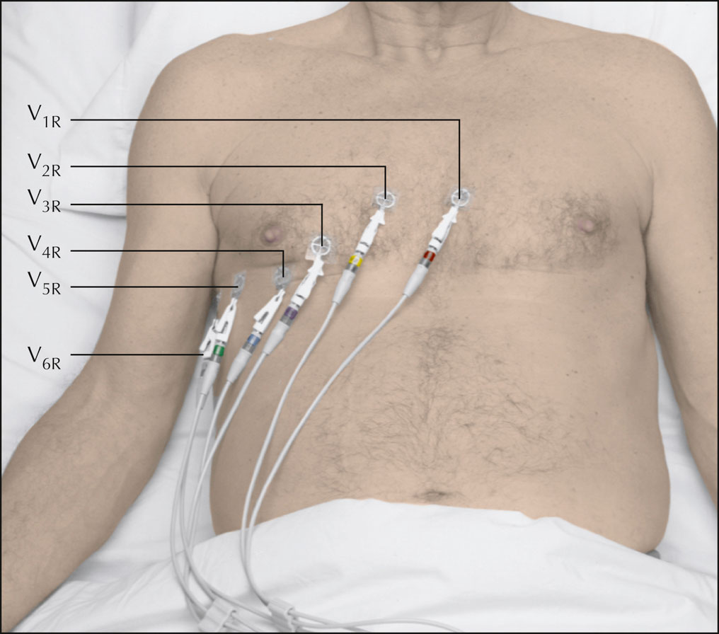
  • Use your fingers to feel between the patient’s ribs (the intercostal spaces). Start at the second intercostal space on the left (the notch felt at the top of the sternum, where the manubrium joins the body of the sternum). Count down two spaces to the fourth intercostal space at the left sternal border. Apply a disposable pre-gelled electrode to the site, and attach lead wire V1R to that electrode.2
  • Move your fingers across the sternum to the fourth intercostal space at the right sternal border. Apply a disposable pre-gelled electrode to that site and attach lead V2R.2
  • Move your finger down to the fifth intercostal space and over to the midclavicular line. Place a disposable pre-gelled electrode here and attach lead V4R. Apply a disposable pre-gelled electrode midway on this line from V2R and attach lead V3R.2
  • Move your finger horizontally from V4R to the right midaxillary line. Apply an electrode to this site and attach lead V6R.2
  • Move your fingers along the same horizontal line to the midpoint between V4R and V6R. This is the right anterior midaxillary line. Apply a disposable pre-gelled electrode to this site and attach lead V5R.2
  • Ask the patient to breathe normally and to refrain from talking during the recording so that muscle movement won’t distort the tracing.
  • Make sure that the paper speed is set at 25 mm/second and the amplitude at 1 mV/10 mm.10,11
  • Press the AUTO or START key. The ECG machine will record all 12 leads automatically. Check your facility’s policy for the number of readings to obtain. (Some facilities require at least two ECGs so that one copy can be sent out for interpretation while the other remains at the bedside.)
  • Observe the tracing quality. If any part of the waveform height extends beyond the paper when you record the ECG, adjust the normal standardization to half-standardization. Note this adjustment on the ECG strip, because this will need to be considered in interpreting the results.25
  • When you’re finished recording the ECG, turn off the machine. If the ECG machine hasn’t already done so, label the ECG with the date and time as well as the patient’s name and identification number. Also label the tracing as RIGHT CHEST ECG to distinguish it from a standard 12-lead ECG.1 Make sure that the tracings are correctly labeled V1R through V6R.
  • Remove the electrodes and discard them in the appropriate receptacle.
  • Clean the patient’s skin with a gauze pad or moist cloth.
  • Return the bed to the lowest position to prevent falls and maintain patients safety.26
  • Remove and discard your gloves, if worn.16,19
  • Perform hand hygiene.4,5,6,7,8,9
  • Put on gloves, as needed.19
  • Clean and disinfect the ECG equipment according to the manufacturer’s instructions to prevent the spread of infection.27,28 Prepare the equipment for future use.2
  • Remove and discard your gloves, as needed.19
  • Perform hand hygiene.4,5,6,7,8,9
  • Place the ECG tracing in the patient’s medical record.
  • Document the procedure.29,30,31,32

Special Considerations

Special considerations
  • If your facility has the capability of transmitting the patient’s ECG from the ECG machine to the patient’s medical record, verify that the ECG transmission was successful and complete.
  • If an additional ECG is necessary, place the patient in the same position as previous ECG tracings to ensure that any ECG changes aren’t the result of body position change.2

Complications

Complications

Prolonged application of the adhesive on the electrodes may cause skin irritation or tissue breakdown. Failure to identify the tracing as a right chest ECG could lead to misdiagnosis.2

Documentation

Documentation

Document the date and time of the procedure, the reason for the test, and the patient’s tolerance of it. Document teaching provided to the patient and family (if applicable), their understanding of that teaching, and any need for follow-up teaching.

References

  1. ProvinseJ. F., et al. (2013). ENA’s translation into practice: Right-sided and posterior electrocardiograms (ECGs). https://www.loyolamedicine.org/sites/default/files/gme/internal-medicine/rightsideecg_0.pdf (Level VII)
  2. WiegandD. L. (2017). AACN procedure manual for high acuity, progressive, and critical care (7th ed.). St. Louis, MO: Elsevier.
  3. NagamM. R., et al. (2017). ECG diagnosis:

    Right ventricular myocardial infarction

    . The Permanente Journal, 21, 16105. https://www.ncbi.nlm.nih.gov/pmc/articles/PMC5267627/
  4. The Joint Commission. (2021). Standard NPSG.07.01.01. Comprehensive accreditation manual for hospitals. Oakbrook Terrace, IL: The Joint Commission. (Level VII)
  5. Centers for Disease Control and Prevention. (2002). Guideline for hand hygiene in health-care settings:

    Recommendations of the Healthcare Infection Control Practices Advisory Committee and the HICPAC/SHEA/APIC/IDSA Hand Hygiene Task Force

    . MMWR Recommendations and Reports, 51(RR-16), 145. https://www.cdc.gov/mmwr/pdf/rr/rr5116.pdf (Level II)
  6. World Health Organization. (2009). WHO guidelines on hand hygiene in health care: First global patient safety challenge, clean care is safer care. https://apps.who.int/iris/bitstream/handle/10665/44102/9789241597906_eng.pdf?sequence=1 (Level IV)
  7. Centers for Medicare and Medicaid Services, Department of Health and Human Services. (2019). Condition of participation:

    Infection control. 42 C.F.R. § 482.42

    .
  8. Accreditation Association for Hospitals and Health Systems. (2020). Standard 07.01.21. Healthcare Facilities Accreditation Program:

    Accreditation requirements for acute care hospitals

    . Chicago, IL: Accreditation Association for Hospitals and Health Systems. (Level VII)
  9. DNV GL-Healthcare USA, Inc. (2020). IC.1.SR.1. NIAHO® accreditation requirements, interpretive guidelines and surveyor guidance-revision 20.0. Milford, OH: DNV GL-Healthcare USA, Inc. (Level VII)
  10. The Joint Commission. (2021). Standard NPSG.01.01.01. Comprehensive accreditation manual for hospitals. Oakbrook Terrace, IL: The Joint Commission. (Level VII)
  11. Centers for Medicare and Medicaid Services, Department of Health and Human Services. (2020). Condition of participation:

    Patient’s rights. 42 C.F.R. § 482.13(c)(1)

    .
  12. Accreditation Association for Hospitals and Health Systems. (2020). Standard 15.01.16. Healthcare Facilities Accreditation Program:

    Accreditation requirements for acute care hospitals

    . Chicago, IL: Accreditation Association for Hospitals and Health Systems. (Level VII)
  13. The Joint Commission. (2021). Standard RI.01.01.01. Comprehensive accreditation manual for hospitals. Oakbrook Terrace, IL: The Joint Commission. (Level VII)
  14. DNV GL-Healthcare USA, Inc. (2020). PR.2.SR.5. NIAHO® accreditation requirements, interpretive guidelines and surveyor guidance-revision 20.0. Milford, OH: DNV GL-Healthcare USA. Inc. (Level VII)
  15. The Joint Commission. (2021). Standard PC.02.01.21. Comprehensive accreditation manual for hospitals. Oakbrook Terrace, IL: The Joint Commission. (Level VII)
  16. SiegelJ. D., et al. (2007, revised 2019). 2007 guideline for isolation precautions: Preventing transmission of infectious agents in healthcare settings. https://www.cdc.gov/infectioncontrol/pdf/guidelines/isolation-guidelines-H.pdf (Level II)
  17. WatersT. R., et al. (2009). Safe patient handling training for schools of nursing. https://www.cdc.gov/niosh/docs/2009-127/pdfs/2009-127.pdf (Level VII)
  18. Accreditation Association for Hospitals and Health Systems. (2020). Standard 07.01.10. Healthcare Facilities Accreditation Program:

    Accreditation requirements for acute care hospitals

    . Chicago, IL: Accreditation Association for Hospitals and Health Systems. (Level VII)
  19. Occupational Safety and Health Administration. (2012). Bloodborne pathogens, standard number 1910.1030. https://www.osha.gov/pls/oshaweb/owadisp.show_document?p_id=10051&p_table=STANDARDS (Level VII)
  20. KligfieldP., et al. (2007). Recommendations for the standardization and interpretation of the electrocardiogram:

    Part I: The electrocardiogram and its technology

    . Circulation, 115(10), 13061324. https://www.ahajournals.org/doi/full/10.1161/circulationaha.106.180200 (Level VII)
  21. OsterC. D. (2005). Proper skin prep helps ensure ECG trace quality. http://multimedia.3m.com/mws/media/358372O/proper-skin-prep-ecg-trace-quality-white-paper.pdf
  22. American Association of Critical-Care Nurses. (2018). AACN practice alert: Managing alarms in acute care across the life span: Electrocardiography and pulse oximetry. https://www.aacn.org/clinical-resources/practice-alerts/managing-alarms-in-acute-care-across-the-life-span (Level VII)
  23. American Association of Critical-Care Nurses. (2018). AACN practice alert: Accurate dysrhythmia monitoring in adults. https://www.aacn.org//clinical-resources/practice-alerts/dysrhythmia-monitoring (Level VII)
  24. American Association of Critical-Care Nurses. (2018). AACN practice alert: Ensuring accurate ST-segment monitoring. https://www.aacn.org/clinical-resources/practice-alerts/st-segment-monitoring (Level VII)
  25. KadishA. H., et al. (2001). ACC/AHA clinical competence statement on electrocardiography and ambulatory electrocardiography. Circulation, 10(25), 31693178. https://www.ahajournals.org/doi/full/10.1161/circ.104.25.3169 (Level VII)
  26. GanzD. A., et al. (2013, reviewed 2021). Preventing falls in hospitals:

    A toolkit for improving quality of care

    (AHRQ Publication No. 13-0015-EF). https://www.ahrq.gov/professionals/systems/hospital/fallpxtoolkit/index.html (Level VII)
  27. RutalaW. A., et al. (2008, revised 2019). Guideline for disinfection and sterilization in healthcare facilities, 2008. https://www.cdc.gov/infectioncontrol/pdf/guidelines/disinfection-guidelines-H.pdf (Level I)
  28. Accreditation Association for Hospitals and Health Systems. (2020). Standard 07.02.03. Healthcare Facilities Accreditation Program:

    Accreditation requirements for acute care hospitals

    . Chicago, IL: Accreditation Association for Hospitals and Health Systems. (Level VII)
  29. The Joint Commission. (2021). Standard RC.01.03.01. Comprehensive accreditation manual for hospitals. Oakbrook Terrace, IL: The Joint Commission. (Level VII)
  30. Centers for Medicare and Medicaid Services, Department of Health and Human Services. (2020). Condition of participation:

    Medical record services. 42 C.F.R. § 482.24(b)

    .
  31. Accreditation Association for Hospitals and Health Systems. (2020). Standard 10.00.03. Healthcare Facilities Accreditation Program:

    Accreditation requirements for acute care hospitals

    . Chicago, IL: Accreditation Association for Hospitals and Health Systems. (Level VII)
  32. DNV GL-Healthcare USA, Inc. (2020). MR.2.SR.1. NIAHO® accreditation requirements, interpretive guidelines and surveyor guidance-revision 20.0. Milford, OH: DNV GL-Healthcare USA, Inc. (Level VII)