The anterior abdominal wall is outlined superiorly by the lower edge of the ribcage and inferiorly by the iliac crests, inguinal ligaments, and pubic bones.
Layers of the Anterior Abdominal Wall
Skin. This consists of the epidermis and the underlying denser tissue of the dermis.
Subcutaneous layer. This is adipose tissue with intervening fibrous septa. Camper fascia is superficial and fatty. It continues caudally into the mons and labia majora. Scarpa fascia is the deeper, more fibrous aspect and is recognizable as a distinct layer.
Musculo-aponeurotic layer. Deep to the subcutaneous tissue are the layered muscles of the anterior abdominal wall and their aponeuroses.
Oblique flank muscles. Most superficial, the external oblique muscle originates from the lower eight ribs and iliac crest, with fibers traveling anteriorly and inferiorly toward the pubic symphysis. Deep to the external, the internal oblique muscle originates from the anterior two-thirds of the iliac crest, the lateral part of the inguinal ligament, and the thoracolumbar fascia in the lower posterior flank. Fibers travel anteriorly and superiorly toward the xiphoid. The transversus abdominis muscle is deep to the internal oblique. It originates from the lower six costal cartilages, the thoracolumbar fascia, the anterior three-fourths of the iliac crest, and the lateral inguinal ligament. Fibers travel transversely.
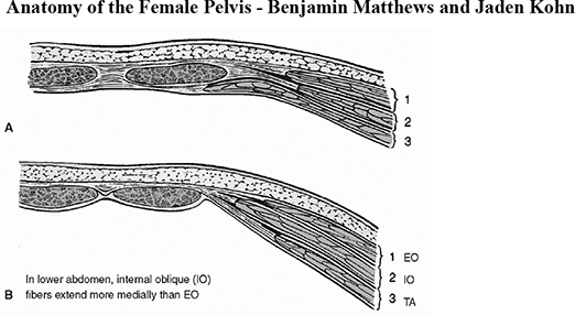
The rectus abdominis muscle is a paired, longitudinal muscle found on either side of the midline, joined medially at the linea alba. The rectus muscle originates from the sternum and cartilage of ribs 5 to 7 and inserts on the anterior surface of the pubic bone. The rectus abdominis muscles are enveloped by the rectus sheath, which is comprised of the aponeuroses of the oblique flank muscles. The structure of the rectus sheath differs above and below the arcuate line (linea semicircularis, semilunar fold of Douglas), which occurs midway between the umbilicus and pubic symphysis. See Figure 58-1.
Above the arcuate line, there are distinct anterior and posterior sheaths investing the rectus muscles. The anterior rectus sheath is comprised of the aponeuroses of the external oblique and the ventral half of the internal oblique. The posterior rectus sheath is comprised of the aponeuroses of the dorsal half of the internal oblique and the transversus abdominis muscles.
Below the arcuate line, there is not a distinct posterior sheath. The anterior sheath is comprised of the aponeuroses of all three oblique flank muscles.
Figure 58-1 Layers of the Abdominal Wall.
A: Cephalad to the arcuate line. B: Caudal to the arcuate line. 1: EO—external oblique, 2: IO—internal oblique, 3: TA—transverse abdominus.
(Reprinted with permission from Corton MM, DeLancey JOL. Surgical anatomy of the female pelvis. In: Van Le L, Handa VL, eds. Te Linde's Operative Gynecology. 13th ed. Wolters Kluwer; 2023:6. Figure 1.4.)
The pyramidalis muscle is a vestigial muscle with variable prominence. It lies superficial to the rectus abdominis muscle, arising from the pubic bone and inserting on the linea alba, several centimeters cephalad to the symphysis pubis.
The transversalis fascia is a fibrous layer, located deep to the abdominal wall muscles. The transversalis is separated from the peritoneum by a variable layer of adipose.
Peritoneum. A single layer of serosa that lines the abdominal wall (parietal peritoneum) and covers the visceral organs (visceral peritoneum). Five vertical peritoneal folds converge toward the umbilicus. The median umbilical fold is a single, midline fold created by the obliterated urachus (which connected the bladder to the umbilicus). The dome of the bladder rises superiorly in the midline and blends into this structure. The medial umbilical folds or ligaments are paired structures lateral to the midline that are now-obliterated remnants of the umbilical arteries. They can be traced back to their origins off the anterior division of the internal iliac artery. The lateral umbilical folds are even more lateral and are formed by the underlying inferior epigastric vessels.
Vasculature of the Abdominal Wall
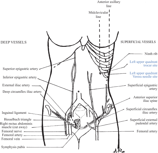
Subcutaneous vascular supply (Fig. 58-2). The subcutaneous tissue is perfused by vessels arising from the femoral artery. The superficial epigastric artery branches off near the femoral canal and runs cephalad approximately 5 cm lateral to the midline. The superficial circumflex iliac artery typically branches off more distally, perfuses the inguinal region, and then runs superolaterally toward the flank.
Figure 58-2 Vasculature and Laparoscopic Landmarks of the Anterior Abdominal Wall.
(Original drawing by Alice W. Ko. From The Johns Hopkins Manual of Gynecology and Obstetrics. 2nd ed. Lippincott Williams and Wilkins; 2002.)
Musculofascial blood supply parallels the subcutaneous supply. The muscles and fascia of the inferior abdominal wall are perfused by branches of the external iliac artery before it traverses the femoral canal. The inferior epigastric artery runs cephalad in a plane deep to the transversalis fascia until the arcuate line, where it then enters a plane between the dorsal belly of the rectus muscle and the posterior rectus sheath. The vessel courses medially from ∼8 cm off midline at its origin (near the deep inguinal ring) to ∼4 cm near the umbilicus. Numerous branches (“perforators”) supply all layers of the abdominal wall before the vessel anastomoses cephalad with the superior epigastric vessels. The deep circumflex iliac artery also branches from the external iliac artery and runs toward the flank between the internal oblique and transversus abdominis muscles.