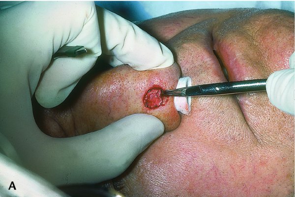
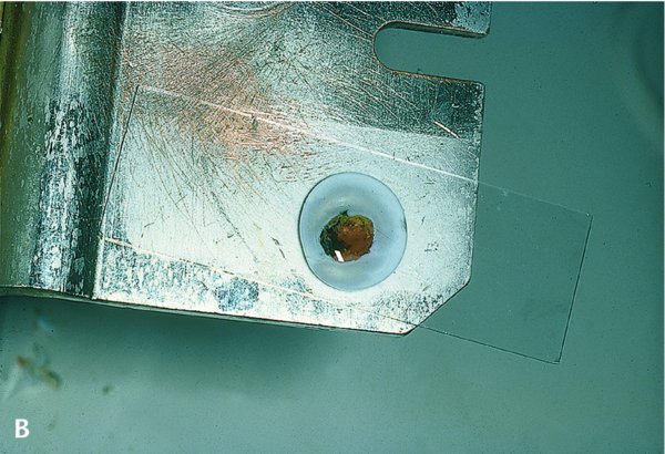
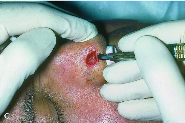
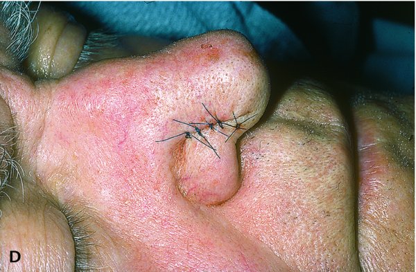
Mohs micrographic surgery.


A:First stage of excision of lesion.
B:Excised tissue color-coded, then evacuated by frozen section.
C:Second stage of excision because the first had positive margins.
D:Primary closure after the second stage was free of malignancy.
(Images courtesy of Michael J. Mulvaney, MD.)