section name header

Disease Prologue

Summary

Cancers Staged Using This Staging System

All carcinomas of the vulva

Cancers Not Staged Using This Staging System

These histopathologic types of cancer…Are staged according to the classification for…and can be found in chapter…
Melanoma of the vulvaMelanoma of the skin47
Extraskeletal Ewing sarcomaSoft Tissue Sarcoma of the Abdomen and Thoracic Visceral Organs42

Summary of Changes

ChangeDetails of ChangeLevel of Evidence
Histopathologic TypeMelanoma was removed from this chapter and is considered separately (Chapter 47).I
Histopathologic TypeClassification of p16 status will be included if obtained but is not requiredIII

ICD-O-3 Topography Codes

CodeDescription
C51.0Labium majus
C51.1Labium minus
C51.2Clitoris
C51.8Overlapping lesion of vulva
C51.9Vulva, NOS

WHO Classification of Tumors

This list includes histology codes and preferred terms from the WHO Classification of Tumors and the International Classification of Diseases for Oncology (ICD-O). Most of the terms in this list represent malignant behavior. For cancer reporting purposes, behavior codes /3 (denoting malignant neoplasms), /2 (denoting in situ neoplasms), and in some cases /1 (denoting neoplasms with uncertain and unknown behavior) may be appended to the 4-digit histology codes to create a complete morphology code.

CodeDescription
8000Neoplasm, malignant
8010Carcinoma, NOS
8010Carcinoma in situ, NOS
8013Large cell neuroendocrine carcinoma
8051Squamous cell carcinoma, verrucous
8051Squamous cell carcinoma, warty
8070Squamous cell carcinoma
8071Squamous cell carcinoma, keratinizing
8072Squamous cell carcinoma, non-keratinizing
8076Squamous cell carcinoma, micro invasive
8077High-grade squamous intraepithelial neoplasia
8083Squamous cell carcinoma, basaloid
8090Basal cell carcinoma
8097Basal cell carcinoma, nodular
8120Transitional cell carcinoma
8140Adenocarcinoma
8140Adenocarcinoma of intestinal type
8140Adenocarcinoma of Skene gland origin
8140Adenocarcinoma of sweat gland type
8200Adenoid cystic carcinoma
8500Adenocarcinoma of mammary gland type
8542Paget disease
8560Adenosquamous carcinoma
9020Phyllodes tumor, malignant
9071Yolk sac tumor

Histology is not ideal for clinical use in patient care, as it describes an unspecified or outdated diagnosis. Data collectors may use this code only if there is not enough information in the medical record to document a more specific diagnosis.

Kurman RJ, Carcangiu ML, Herrington CS, Young RH, eds. World Health Organization Classification of Tumours of the Female Reproductive System. Lyon: IARC; 2014.

International Agency for Research on Cancer, World Health Organization. International Classification of Diseases for Oncology. ICD-O-3-Online.http://codes.iarc.fr/home. Accessed August 16, 2017. Used with permission.

Introduction

The classification system for vulvar cancers has been modified for the AJCC Cancer Staging Manual, 8 th Edition of TNM in accordance with changes adopted by the Federation Internationale de Gynecologie et d'Obstetrique (FIGO) to remove melanoma as a histologic type for vulvar cancer and to classify it as though it were a cutaneous melanoma, and to further help delineate the appropriate anatomic classification of anterior perineal lesions.

Anatomy

Primary Site(s)

The vulva is the anatomic area immediately external to the vagina. It includes the labia, clitoris, and perineum. The tumor may extend to involve the vagina, urethra, or anus (Figure 50.1). It may be fixed to the pubic bone. Changes to the staging classification reflect a belief that tumor size independent of other factors (spread to adjacent structures, nodal metastases) is less important in predicting survival.

50.1 Vulva and perineum lesions, from top to bottom: the lesion at the top is vulvar, the middle two lesions are perineal, and the lesion at the bottom is considered perianal.

Regional Lymph Nodes

The femoral and inguinal nodes are the sites of regional spread (Figure 50.2).

50.2 Regional lymph nodes of the vulva.

Metastatic Sites

The metastatic sites include any site beyond the area of the regional lymph nodes. Tumor involvement of pelvic lymph nodes, including internal iliac, external iliac, and common iliac lymph nodes, is considered distant metastasis.

Classification Rules

Clinical Classification

Cases should be classified as carcinoma of the vulva if the primary site of the growth is in the vulva. Tumors present on the vulva as secondary growths from either a genital or an extragenital site should be excluded. There should be histologic confirmation of the tumor.

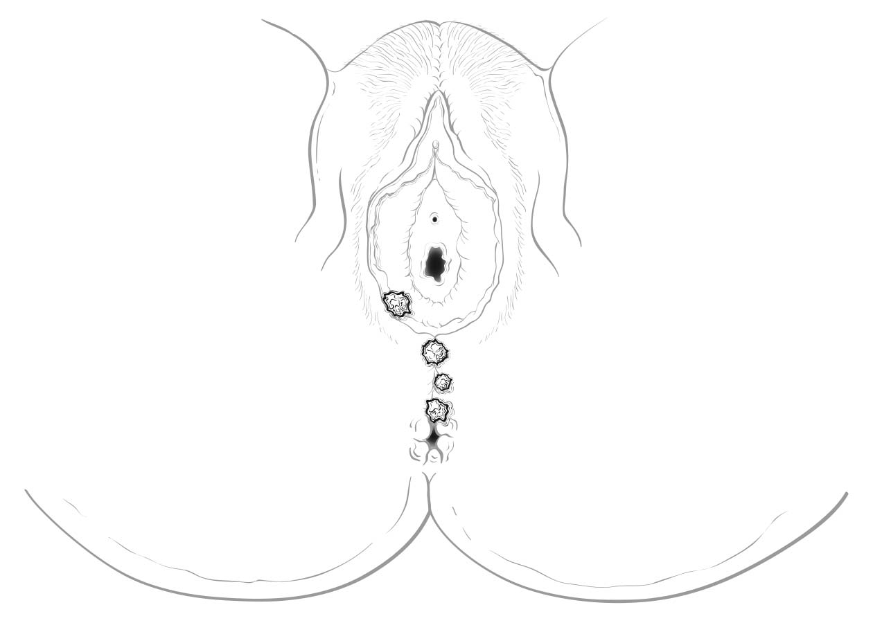
Perineal lesions represent a challenging subset of cancers that may arise from either the vulva or the perianal mucosa. Anterior perineal lesions may be considered either vulvar or perianal, and the treatment plans may be quite dissimilar. For this reason, we recommend the following: Lesions that clearly arise from the vulva and extend onto the perineum and potentially involve the anus should be classified as vulvar. Similarly, lesions that clearly arise from the anus and extend onto the perineum should be classified as perianal. For lesions localized to the perineum that do not clearly arise from either the vulva or the anus, we recommend that the clinician denote their exact location as well as his or her favored impression regarding classification. We recommend the following terminology: “perineum favor vulva” and “perineum favor anus.” We also recommend consultation with colleagues in gynecologic oncology and colorectal, general, or surgical oncology, as classification has a significant impact on treatment.

Rarely, lymph nodes are assessed by radiologic-guided fine-needle aspiration or by using imaging techniques such as computed tomography (CT), magnetic resonance (MR) imaging, or positron emission tomography (PET).

Single tumor cells or small clusters of cells not more than 0.2 mm in greatest diameter are classified as isolated tumor cells. These cells may be detected by routine histology or by immunohistochemical methods. They are designated N0(i+).

Imaging

MR imaging, CT, and PET/CT have been used in the staging of vulvar cancer. MR imaging, with its high soft tissue resolution, can stage the disease locally. CT and PET/CT may be used to assess for lymph node metastases and distant disease. Some reports suggest that PET/CT has greater sensitivity in assessing lymph node metastases in vulvar cancer. 1

TNM Components of Tumor Staging

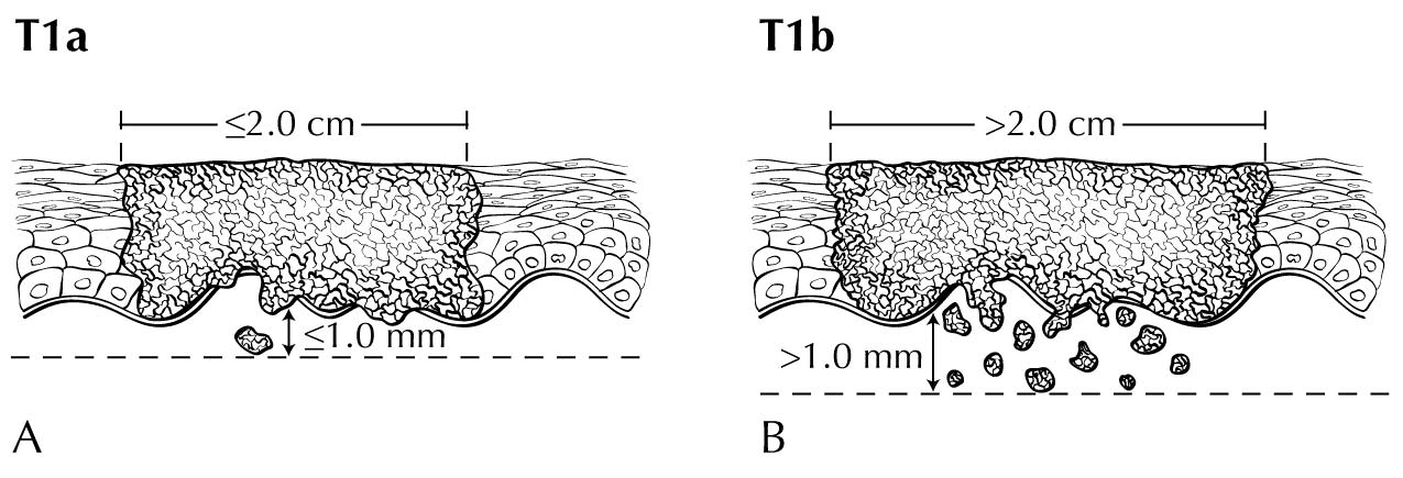
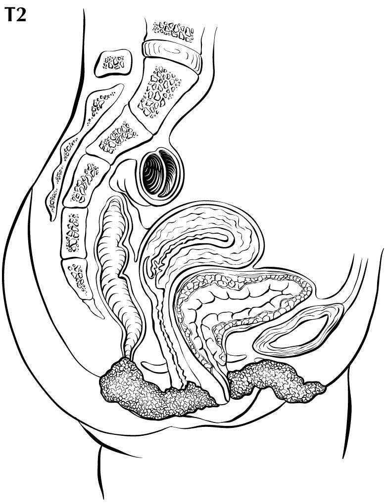
In stage T1 disease, multifocal lesions may be present. Currently, MR imaging is not used to assess the depth of invasion of the tumor into the dermal papilla. Assessing for depth of invasion of 1 mm is beyond the resolution of any currently available imaging modality. However, MR imaging may be used to measure a tumor's size, given its soft tissue resolution. If the tumor is 2 cm or smaller, it is category T1a. The disease may be visualized easier if vaginal gel is instilled and axial dynamic T1-weighted images are obtained. If the tumor is larger than 2 cm, it is category T1b. In category T2 disease, the tumor extends to the lower third of the vagina/urethra and the anus; it may be seen best on MR T2-weighted and dynamic sagittal T1-weighted sequences. In category T3 disease, the tumor may be any size and extends to the upper proximal two thirds of the urethra, the upper proximal two thirds of the vagina, the bladder mucosa, or the rectal mucosa, or it is fixed to pelvic bone. Skeletal involvement is seen best on MR non-fat-saturated T1-weighted sequences. However, involvement of other organs may be seen on MR sagittal T2-weighted and dynamic sagittal T1-weighted sequences.

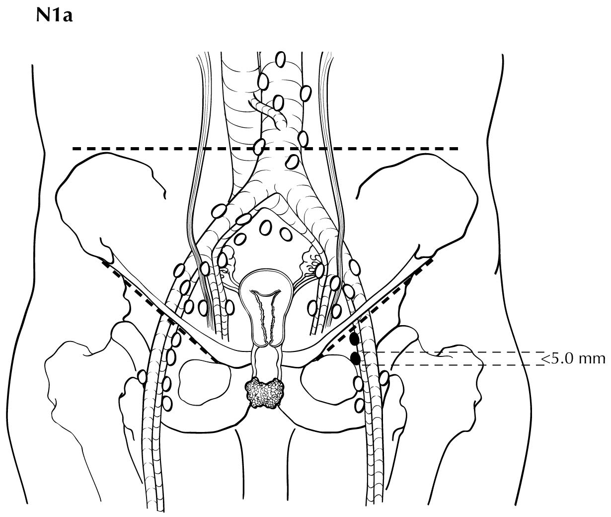
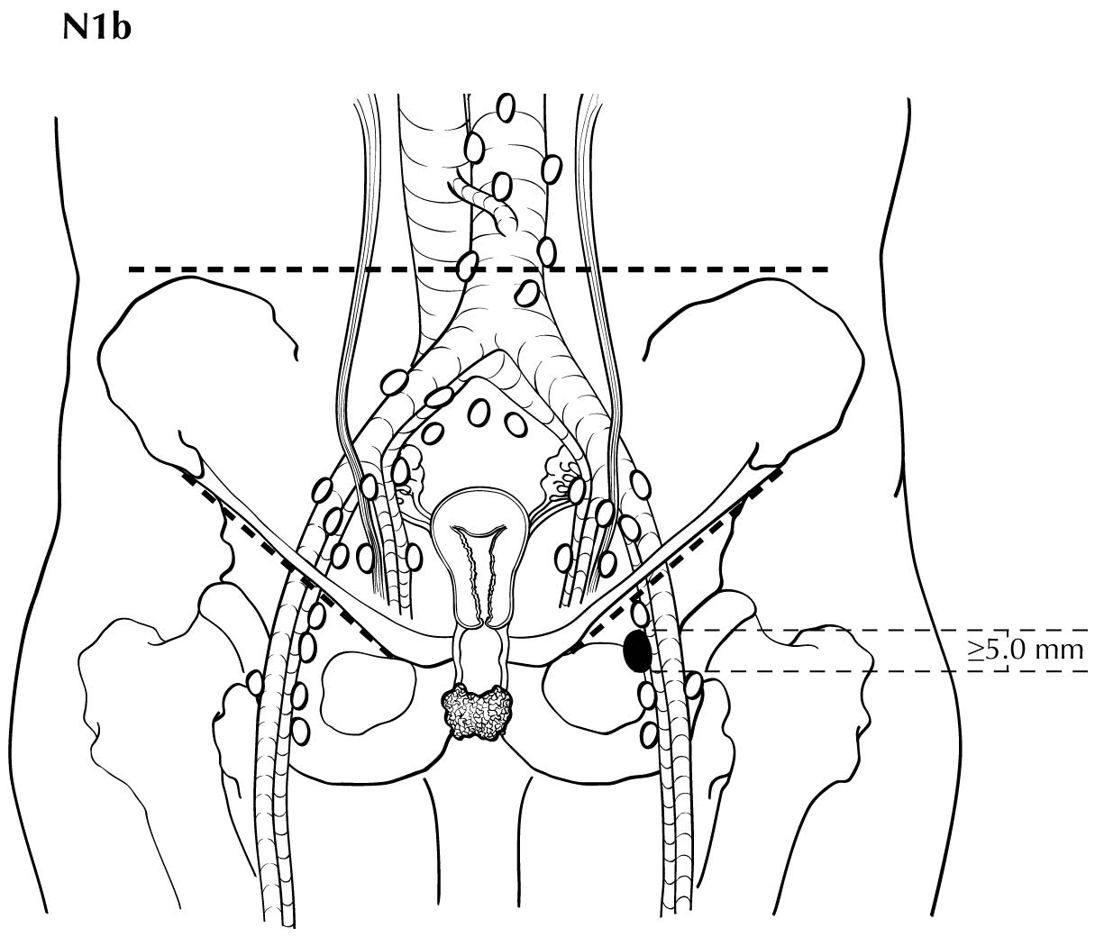
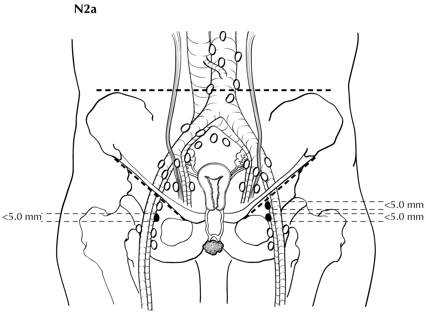
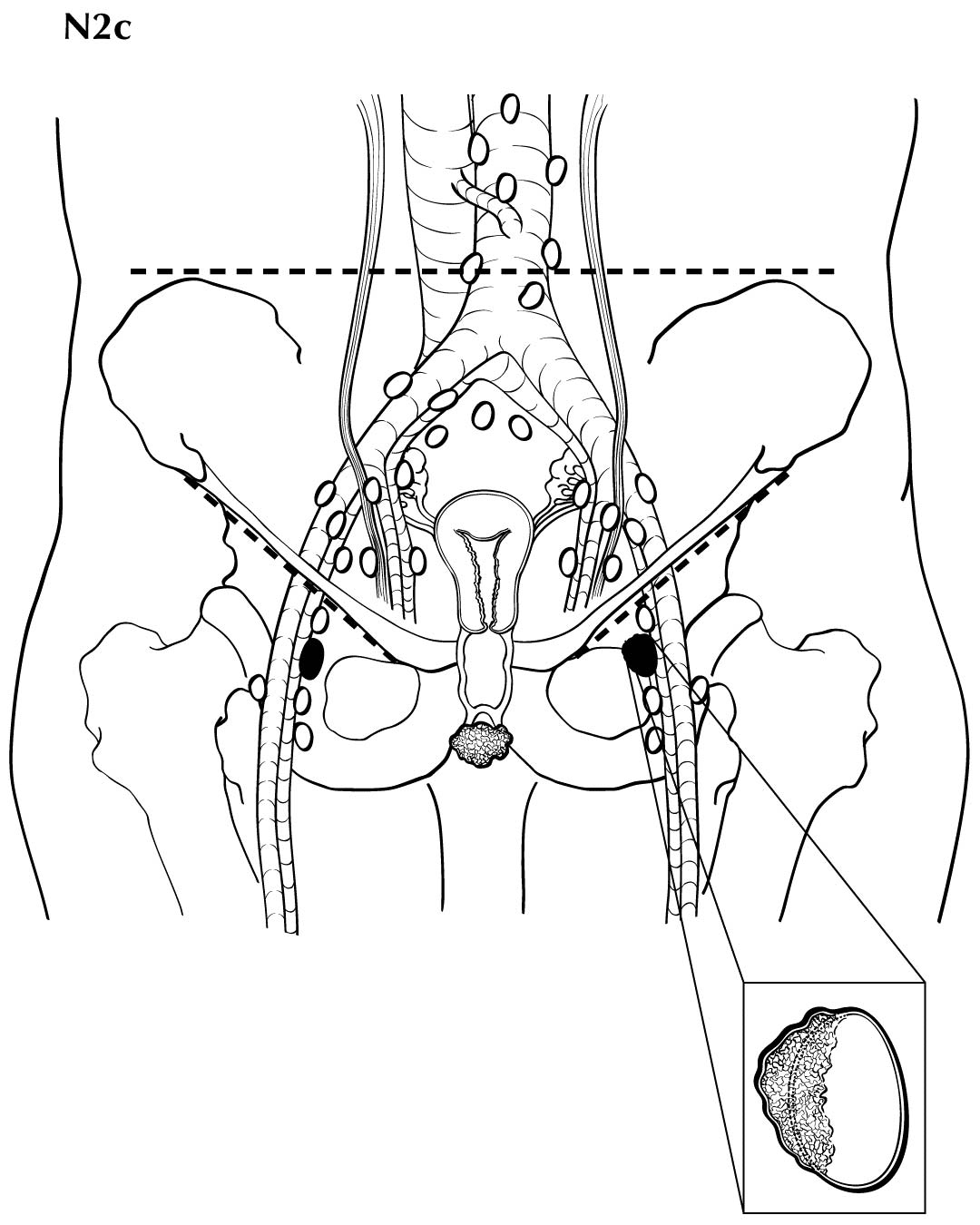
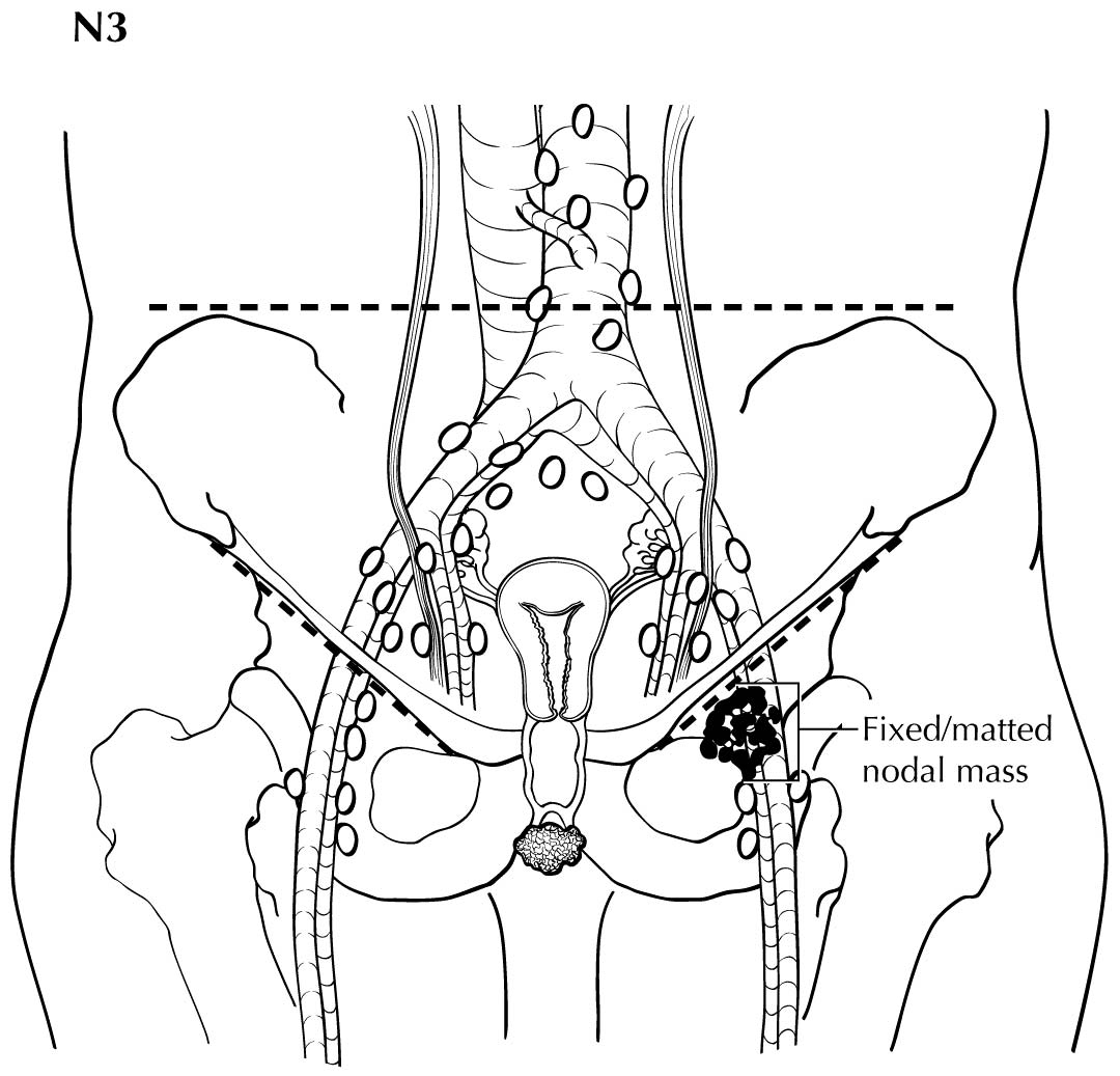
Regional nodal metastases less than 5 mm (histologically) are considered N1 disease and cannot be assessed by cross-sectional imaging. Metastatic disease less than 5 mm is beyond the scope of any in vivo imaging modality. However, some N2 disease can be identified easily based on the number and size of nodes involved. The imaging criteria used to assess lymph node metastases is based on lymph node size, with abnormal being greater than 1 cm in the short axial dimension. However, because there may be false positive causes of enlarged nodes from benign disease, PET/CT is considered superior in assessing lymph node metastases. Metabolically active lymph nodes of any size on PET/CT are considered metastatic. Ulceration and immobility of the lymph nodes may be assessed better on physical examination.

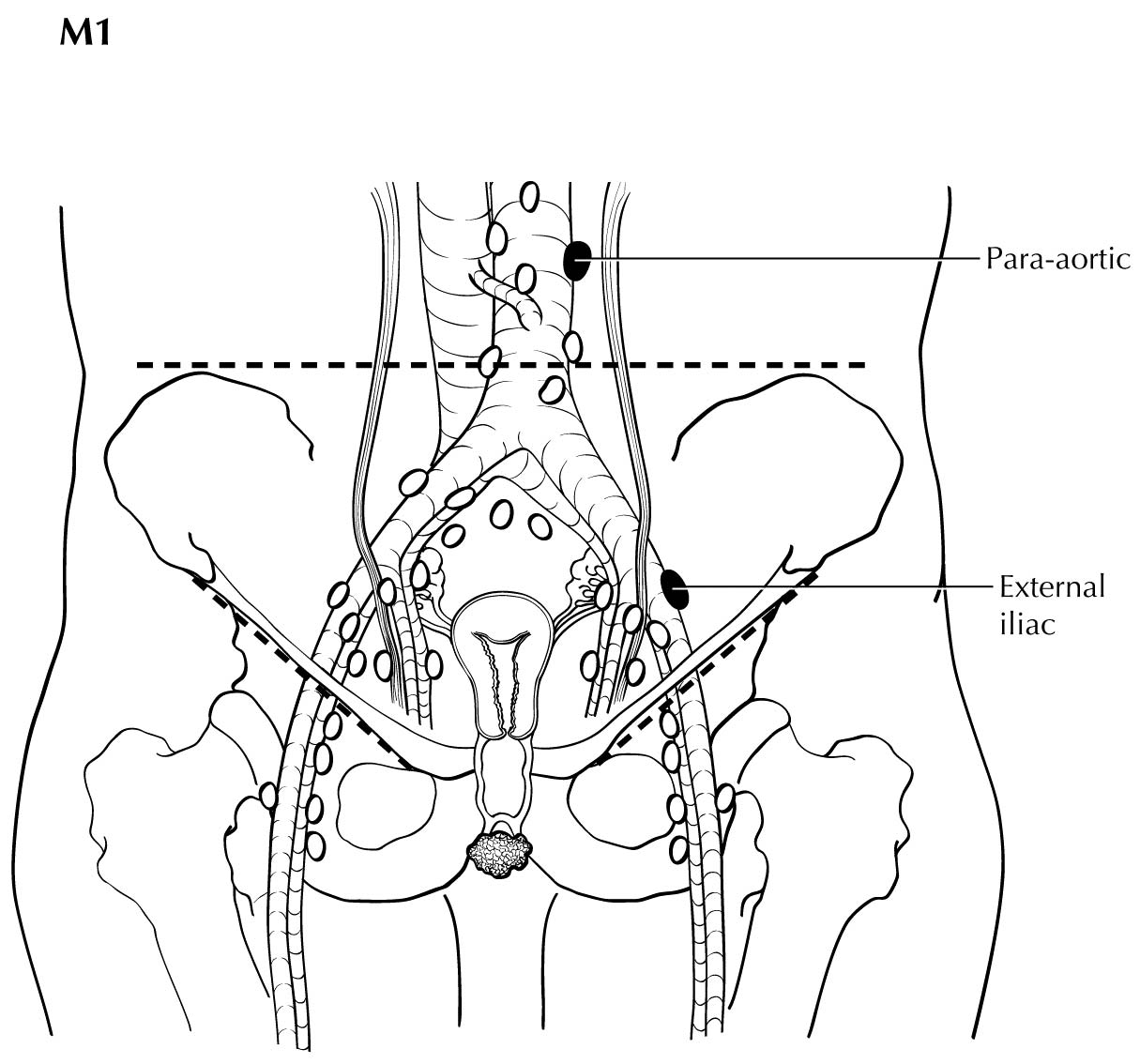
The presence of pelvic lymph node metastases or lung, visceral, or bone metastases is considered M1 disease, and PET/CT is superior in assessing its extent. If PET/CT is not available, contrast-enhanced CT may be used.

Suggested Imaging Report Format

  1. Primary tumor
    1. Size less than or equal to 2 cm or greater than 2 cm
  2. Local extent
    1. Involvement of the bladder and the rectum, vagina, and urethra
    2. Description of the site, number, and laterality of inguinal lymph nodes
  3. Pelvic and distant lymph node involvement and extrapelvic disease

Pathological Classification

FIGO uses surgical/pathological staging for vulvar cancer. Stage should be assigned at the time of definitive surgical treatment before radiation or chemotherapy. If chemotherapy, radiation, or a combination of both treatment modalities is the initial mode of therapy, clinical staging should be used. The stage cannot be changed on the basis of disease progression or recurrence or on the basis of response to initial radiation or chemotherapy that precedes primary tumor resection.

For pN, histologic examination of regional lymphadenectomy specimens ordinarily includes six or more lymph nodes. For TNM staging, cases with fewer than six resected nodes should be classified using the TNM pathological classification based on the status of those nodes (e.g., pN0, pN1) according to the general rules of TNM. The number of resected and positive nodes should be recorded (note that FIGO classifies cases with less than six nodes resected as pNX). The concept of sentinel lymph node mapping, in which only one or two key nodes are removed, currently is being investigated. In most cases, regional lymph nodes are assessed surgically (via inguinal-femoral lymphadenectomy). Included in the 8 th Edition is the opportunity to denote a micrometastatic lymph node using the N1mi or N2mi category. The current revisions to staging adopted reflect the recognition that the number and size of lymph node metastases more accurately reflect prognosis.

Prognostic Factors

Prognostic Factors Required for Stage Grouping

Beyond the factors used to assign T, N, or M categories, no additional prognostic factors are required for stage grouping.

Additional Factors Recommended for Clinical Care

Vulvar cancer is a surgically staged malignancy. Surgical/pathological staging provides specific information about primary tumor size and lymph node status, which are the most important prognostic factors in vulvar cancer. Other commonly evaluated items, such as histologic type, differentiation, DNA ploidy, and S-phase fraction analysis, as well as age, are not identified uniformly as important prognostic factors in vulvar cancer.

FIGO Stage

FIGO stage should be recorded.

AJCC Level of Evidence: I
Femoral-Inguinal Nodal Status and Method of Assessment

Nodal spread identified on MR imaging, CT, or PET is prognostic and should be recorded and used in treatment planning.

AJCC Level of Evidence: I
Pelvic Nodal Status and Method of Assessment

Nodal spread identified on MR imaging, CT, or PET is prognostic and should be recorded and used in treatment planning.

AJCC Level of Evidence: I
p16

Essentially two pathways have been identified in the pathogenesis of invasive vulvar carcinoma. The first pathway is the classic progression of vulvar intraepithelial neoplasia (VIN), which is associated with high-grade human papillomavirus (HPV) infection (commonly HPV subtypes 16 and 18). The second pathway is referred to as differentiated VIN simplex, which is not associated with HPV infection but rather with vulvar dystrophy. In the classic presentation, the VIN tends to be multifocal and is more common in younger women, with a relatively low risk of progression into invasive squamous cell carcinoma of the vulva. It is diffusely positive for p16, a molecular marker that reflects the integration of the HPV genome into the cell. Some centers perform p16 staining on invasive vulvar carcinomas, and that information should be reported and collected for analysis to determine whether the presence of p16 might be used as a prognostic molecular marker in the future.

AJCC Level of Evidence: III

The authors have not noted any emerging factors for clinical care.

Risk Assessment

Risk Assesment Models

The AJCC recently established guidelines that will be used to evaluate published statistical prediction models for the purpose of granting endorsement for clinical use.2 Although this is a monumental step toward the goal of precision medicine, this work was published only very recently. Therefore, the existing models that have been published or may be in clinical use have not yet been evaluated for this cancer site by the Precision Medicine Core of the AJCC. In the future, the statistical prediction models for this cancer site will be evaluated, and those that meet all AJCC criteria will be endorsed.

Recommendations

The authors have not provided recommendations for clinical trial stratification.

TNM Definitions

Clinical T (cT)

cT CategoryFIGO StagecT Criteria
cTXN/APrimary tumor cannot be assessed
cT0N/ANo evidence of primary tumor
cT1ITumor confined to the vulva and /or perineum. Multifocal lesions should be designated as such. The largest lesion or the lesion with the greatest depth of invasion will be the target lesion identified to address the highest pT stage. Depth of invasion is defined as the measurement of the tumor from the epithelial-stromal junction of the adjacent most superficial dermal papilla to the deepest point of invasion.
cT1aIALesions 2 cm or less, confined to the vulva and /or perineum, and with stromal invasion of 1.0 mm or less
cT1bIBLesions more than 2 cm, or any size with stromal invasion of more than 1.0 mm, confined to the vulva and /or perineum
cT2IITumor of any size with extension to adjacent perineal structures (lower/distal third of the urethra, lower/distal third of the vagina, anal involvement)
cT3IVATumor of any size with extension to any of the following—upper/proximal two thirds of the urethra, upper/proximal two thirds of the vagina, bladder mucosa, or rectal mucosa—or fixed to pelvic bone

Pathological T (pT)

pT CategoryFIGO StagepT Criteria
pTXN/APrimary tumor cannot be assessed
pT0N/ANo evidence of primary tumor
pT1ITumor confined to the vulva and /or perineum. Multifocal lesions should be designated as such. The largest lesion or the lesion with the greatest depth of invasion will be the target lesion identified to address the highest pT stage. Depth of invasion is defined as the measurement of the tumor from the epithelial-stromal junction of the adjacent most superficial dermal papilla to the deepest point of invasion.
pT1aIALesions 2 cm or less, confined to the vulva and /or perineum, and with stromal invasion of 1.0 mm or less
pT1bIBLesions more than 2 cm, or any size with stromal invasion of more than 1.0 mm, confined to the vulva and /or perineum
pT2IITumor of any size with extension to adjacent perineal structures (lower/distal third of the urethra, lower/distal third of the vagina, anal involvement)
pT3IVATumor of any size with extension to any of the following—upper/proximal two thirds of the urethra, upper/proximal two thirds of the vagina, bladder mucosa, or rectal mucosa—or fixed to pelvic bone
cTXN/APrimary tumor cannot be assessed
cT0N/ANo evidence of primary tumor
cT1ITumor confined to the vulva and /or perineum. Multifocal lesions should be designated as such. The largest lesion or the lesion with the greatest depth of invasion will be the target lesion identified to address the highest pT stage. Depth of invasion is defined as the measurement of the tumor from the epithelial-stromal junction of the adjacent most superficial dermal papilla to the deepest point of invasion.
cT1aIALesions 2 cm or less, confined to the vulva and /or perineum, and with stromal invasion of 1.0 mm or less
cT1bIBLesions more than 2 cm, or any size with stromal invasion of more than 1.0 mm, confined to the vulva and /or perineum
cT2IITumor of any size with extension to adjacent perineal structures (lower/distal third of the urethra, lower/distal third of the vagina, anal involvement)
cT3IVATumor of any size with extension to any of the following—upper/proximal two thirds of the urethra, upper/proximal two thirds of the vagina, bladder mucosa, or rectal mucosa—or fixed to pelvic bone

Neoadjuvant Clinical T (yT)

ycT CategoryFIGO StageycT Criteria
ycTXN/APrimary tumor cannot be assessed
ycT0N/ANo evidence of primary tumor
ycT1ITumor confined to the vulva and /or perineum. Multifocal lesions should be designated as such. The largest lesion or the lesion with the greatest depth of invasion will be the target lesion identified to address the highest pT stage. Depth of invasion is defined as the measurement of the tumor from the epithelial-stromal junction of the adjacent most superficial dermal papilla to the deepest point of invasion.
ycT1aIALesions 2 cm or less, confined to the vulva and /or perineum, and with stromal invasion of 1.0 mm or less
ycT1bIBLesions more than 2 cm, or any size with stromal invasion of more than 1.0 mm, confined to the vulva and /or perineum
ycT2IITumor of any size with extension to adjacent perineal structures (lower/distal third of the urethra, lower/distal third of the vagina, anal involvement)
ycT3IVATumor of any size with extension to any of the following—upper/proximal two thirds of the urethra, upper/proximal two thirds of the vagina, bladder mucosa, or rectal mucosa—or fixed to pelvic bone

Neoadjuvant Pathological T (yT)

ypT CategoryFIGO StageypT Criteria
ypTXN/APrimary tumor cannot be assessed
ypT0N/ANo evidence of primary tumor
ypT1ITumor confined to the vulva and /or perineum. Multifocal lesions should be designated as such. The largest lesion or the lesion with the greatest depth of invasion will be the target lesion identified to address the highest pT stage. Depth of invasion is defined as the measurement of the tumor from the epithelial-stromal junction of the adjacent most superficial dermal papilla to the deepest point of invasion.
ypT1aIALesions 2 cm or less, confined to the vulva and /or perineum, and with stromal invasion of 1.0 mm or less
ypT1bIBLesions more than 2 cm, or any size with stromal invasion of more than 1.0 mm, confined to the vulva and /or perineum
ypT2IITumor of any size with extension to adjacent perineal structures (lower/distal third of the urethra, lower/distal third of the vagina, anal involvement)
ypT3IVATumor of any size with extension to any of the following—upper/proximal two thirds of the urethra, upper/proximal two thirds of the vagina, bladder mucosa, or rectal mucosa—or fixed to pelvic bone
Pathological N (pN)
pN CategoryFIGO StagepN Criteria
pNXN/ARegional lymph nodes cannot be assessed
pN0N/ANo regional lymph node metastasis
pN0(i+)N/AIsolated tumor cells in regional lymph node(s) no greater than 0.2 mm
pN1IIIRegional lymph node metastasis with one or two lymph node metastases each less than 5 mm, or one lymph node metastasis greater than or equal to 5 mm
pN1aIIIAOne or two lymph node metastases each less than 5 mm
pN1bIIIAOne lymph node metastasis greater than or equal to 5 mm
pN2N/ARegional lymph node metastasis with three or more lymph node metastases each less than 5 mm, or two or more lymph node metastases greater than or equal to 5 mm, or lymph node(s) with extranodal extension
pN2aIIIBThree or more lymph node metastases each less than 5 mm
pN2bIIIBTwo or more lymph node metastases greater than or equal to 5 mm
pN2cIIICLymph node(s) with extranodal extension
pN3IVAFixed or ulcerated regional lymph node metastasis
cNXN/ARegional lymph nodes cannot be assessed
cN0N/ANo regional lymph node metastasis
cN0(i+)N/AIsolated tumor cells in regional lymph node(s) no greater than 0.2 mm
cN1IIIRegional lymph node metastasis with one or two lymph node metastases each less than 5 mm, or one lymph node metastasis greater than or equal to 5 mm
cN1aIIIAOne or two lymph node metastases each less than 5 mm
cN1bIIIAOne lymph node metastasis greater than or equal to 5 mm
cN2N/ARegional lymph node metastasis with three or more lymph node metastases each less than 5 mm, or two or more lymph node metastases greater than or equal to 5 mm, or lymph node(s) with extranodal extension
cN2aIIIBThree or more lymph node metastases each less than 5 mm
cN2bIIIBTwo or more lymph node metastases greater than or equal to 5 mm
cN2cIIICLymph node(s) with extranodal extension
cN3IVAFixed or ulcerated regional lymph node metastasis

Includes micrometastasis, N1mi and N2mi.

The site, size, and laterality of lymph node metastasis should be recorded.

Definition of Distant Metastasis (M)- Clinical M (cN)

cM CategoryFIGO StagecM Criteria
cM0N/ANo distant metastasis (no pathological M0; use clinical M to complete stage group)
cM1IVBDistant metastasis (including pelvic lymph node metastasis)
pM1IVBMicroscopic evidence of distant metastasis (including pelvic lymph node metastasis)

Definition of Distant Metastasis (M)- Pathological M (pN)

pM CategoryFIGO StagePM Criteria
cM0N/ANo distant metastasis (no pathological M0; use clinical M to complete stage group)
cM1IVBDistant metastasis (including pelvic lymph node metastasis)
pM1IVBMicroscopic evidence of distant metastasis (including pelvic lymph node metastasis)

Definition of Distant Metastasis (M)- Neoadjuvant Clinical M (pY)

ycM CategoryFIGO StageycM Criteria
cM0N/ANo distant metastasis (no pathological M0; use clinical M to complete stage group)
cM1IVBDistant metastasis (including pelvic lymph node metastasis)
pM1IVBMicroscopic evidence of distant metastasis (including pelvic lymph node metastasis)

Definition of Distant Metastasis (M)- Neoadjuvant Pathological M (pY)

ypM CategoryFIGO StageM Criteria
cM0N/ADistant metastasis (including pelvic lymph node metastasis), microscopically confirmed
cM1IVBNo distant metastasis (no pathological M0; use clinical M to complete stage group)
pM1IVBMicroscopic evidence of distant metastasis (including pelvic lymph node metastasis)

Stage Prognostic

Clinical

When T is…and N is…and M is…Then the Clinical Prognostic Stage Group is…
cT1cN0cM0I
cT1acN0cM0IA
cT1bcN0cM0IB
cT2cN0cM0II
cT1cN0(i+)cM0I
cT1acN0(i+)cM0IA
cT1bcN0(i+)cM0IB
cT2cN0(i+)cM0II
cT1cN2cM0III
cT1acN2cM0III
cT1bcN2cM0III
cT2cN2cM0III
cT1cN1cM0IIIA
cT1cN1acM0IIIA
cT1cN1bcM0IIIA
cT1acN1cM0IIIA
cT1acN1acM0IIIA
cT1acN1bcM0IIIA
cT1bcN1cM0IIIA
cT1bcN1acM0IIIA
cT1bcN1bcM0IIIA
cT2cN1cM0IIIA
cT2cN1acM0IIIA
cT2cN1bcM0IIIA
cT1cN2acM0IIIB
cT1cN2bcM0IIIB
cT1acN2acM0IIIB
cT1acN2bcM0IIIB
cT1bcN2acM0IIIB
cT1bcN2bcM0IIIB
cT2cN2acM0IIIB
cT2cN2bcM0IIIB
cT1cN2ccM0IIIC
cT1acN2ccM0IIIC
cT1bcN2ccM0IIIC
cT2cN2ccM0IIIC
cT1cN3cM0IVA
cT1acN3cM0IVA
cT1bcN3cM0IVA
cT2cN3cM0IVA
cT3cNXcM0IVA
cT3cN0cM0IVA
cT3cN0(i+)cM0IVA
cT3cN1cM0IVA
cT3cN1acM0IVA
cT3cN1bcM0IVA
cT3cN2cM0IVA
cT3cN2acM0IVA
cT3cN2bcM0IVA
cT3cN2ccM0IVA
cT3cN3cM0IVA
cTXcNXcM1IVB
cTXcN0cM1IVB
cTXcN0(i+)cM1IVB
cTXcN1cM1IVB
cTXcN1acM1IVB
cTXcN1bcM1IVB
cTXcN2cM1IVB
cTXcN2acM1IVB
cTXcN2bcM1IVB
cTXcN2ccM1IVB
cTXcN3cM1IVB
cT0cNXcM1IVB
cT0cN0cM1IVB
cT0cN0(i+)cM1IVB
cT0cN1cM1IVB
cT0cN1acM1IVB
cT0cN1bcM1IVB
cT0cN2cM1IVB
cT0cN2acM1IVB
cT0cN2bcM1IVB
cT0cN2ccM1IVB
cT0cN3cM1IVB
cT1cNXcM1IVB
cT1cN0cM1IVB
cT1cN0(i+)cM1IVB
cT1cN1cM1IVB
cT1cN1acM1IVB
cT1cN1bcM1IVB
cT1cN2cM1IVB
cT1cN2acM1IVB
cT1cN2bcM1IVB
cT1cN2ccM1IVB
cT1cN3cM1IVB
cT1acNXcM1IVB
cT1acN0cM1IVB
cT1acN0(i+)cM1IVB
cT1acN1cM1IVB
cT1acN1acM1IVB
cT1acN1bcM1IVB
cT1acN2cM1IVB
cT1acN2acM1IVB
cT1acN2bcM1IVB
cT1acN2ccM1IVB
cT1acN3cM1IVB
cT1bcNXcM1IVB
cT1bcN0cM1IVB
cT1bcN0(i+)cM1IVB
cT1bcN1cM1IVB
cT1bcN1acM1IVB
cT1bcN1bcM1IVB
cT1bcN2cM1IVB
cT1bcN2acM1IVB
cT1bcN2bcM1IVB
cT1bcN2ccM1IVB
cT1bcN3cM1IVB
cT2cNXcM1IVB
cT2cN0cM1IVB
cT2cN0(i+)cM1IVB
cT2cN1cM1IVB
cT2cN1acM1IVB
cT2cN1bcM1IVB
cT2cN2cM1IVB
cT2cN2acM1IVB
cT2cN2bcM1IVB
cT2cN2ccM1IVB
cT2cN3cM1IVB
cT3cNXcM1IVB
cT3cN0cM1IVB
cT3cN0(i+)cM1IVB
cT3cN1cM1IVB
cT3cN1acM1IVB
cT3cN1bcM1IVB
cT3cN2cM1IVB
cT3cN2acM1IVB
cT3cN2bcM1IVB
cT3cN2ccM1IVB
cT3cN3cM1IVB
cTXcNXpM1IVB
cTXcN0pM1IVB
cTXcN0(i+)pM1IVB
cTXcN1pM1IVB
cTXcN1apM1IVB
cTXcN1bpM1IVB
cTXcN2pM1IVB
cTXcN2apM1IVB
cTXcN2bpM1IVB
cTXcN2cpM1IVB
cTXcN3pM1IVB
cT0cNXpM1IVB
cT0cN0pM1IVB
cT0cN0(i+)pM1IVB
cT0cN1pM1IVB
cT0cN1apM1IVB
cT0cN1bpM1IVB
cT0cN2pM1IVB
cT0cN2apM1IVB
cT0cN2bpM1IVB
cT0cN2cpM1IVB
cT0cN3pM1IVB
cT1cNXpM1IVB
cT1cN0pM1IVB
cT1cN0(i+)pM1IVB
cT1cN1pM1IVB
cT1cN1apM1IVB
cT1cN1bpM1IVB
cT1cN2pM1IVB
cT1cN2apM1IVB
cT1cN2bpM1IVB
cT1cN2cpM1IVB
cT1cN3pM1IVB
cT1acNXpM1IVB
cT1acN0pM1IVB
cT1acN0(i+)pM1IVB
cT1acN1pM1IVB
cT1acN1apM1IVB
cT1acN1bpM1IVB
cT1acN2pM1IVB
cT1acN2apM1IVB
cT1acN2bpM1IVB
cT1acN2cpM1IVB
cT1acN3pM1IVB
cT1bcNXpM1IVB
cT1bcN0pM1IVB
cT1bcN0(i+)pM1IVB
cT1bcN1pM1IVB
cT1bcN1apM1IVB
cT1bcN1bpM1IVB
cT1bcN2pM1IVB
cT1bcN2apM1IVB
cT1bcN2bpM1IVB
cT1bcN2cpM1IVB
cT1bcN3pM1IVB
cT2cNXpM1IVB
cT2cN0pM1IVB
cT2cN0(i+)pM1IVB
cT2cN1pM1IVB
cT2cN1apM1IVB
cT2cN1bpM1IVB
cT2cN2pM1IVB
cT2cN2apM1IVB
cT2cN2bpM1IVB
cT2cN2cpM1IVB
cT2cN3pM1IVB
cT3cNXpM1IVB
cT3cN0pM1IVB
cT3cN0(i+)pM1IVB
cT3cN1pM1IVB
cT3cN1apM1IVB
cT3cN1bpM1IVB
cT3cN2pM1IVB
cT3cN2apM1IVB
cT3cN2bpM1IVB
cT3cN2cpM1IVB
cT3cN3pM1IVB

Pathological

When T is…and N is…and M is…Then the Pathological Prognostic Stage Group is…
pT1pN0cM0I
pT1apN0cM0IA
pT1bpN0cM0IB
pT2pN0cM0II
pT1pN0(i+)cM0I
pT1apN0(i+)cM0IA
pT1bpN0(i+)cM0IB
pT2pN0(i+)cM0II
pT1pN2cM0III
pT1apN2cM0III
pT1bpN2cM0III
pT2pN2cM0III
pT1pN1cM0IIIA
pT1pN1acM0IIIA
pT1pN1bcM0IIIA
pT1apN1cM0IIIA
pT1apN1acM0IIIA
pT1apN1bcM0IIIA
pT1bpN1cM0IIIA
pT1bpN1acM0IIIA
pT1bpN1bcM0IIIA
pT2pN1cM0IIIA
pT2pN1acM0IIIA
pT2pN1bcM0IIIA
pT1pN2acM0IIIB
pT1pN2bcM0IIIB
pT1apN2acM0IIIB
pT1apN2bcM0IIIB
pT1bpN2acM0IIIB
pT1bpN2bcM0IIIB
pT2pN2acM0IIIB
pT2pN2bcM0IIIB
pT1pN2ccM0IIIC
pT1apN2ccM0IIIC
pT1bpN2ccM0IIIC
pT2pN2ccM0IIIC
pT1pN3cM0IVA
pT1apN3cM0IVA
pT1bpN3cM0IVA
pT2pN3cM0IVA
pT3pNXcM0IVA
pT3pN0cM0IVA
pT3pN0(i+)cM0IVA
pT3pN1cM0IVA
pT3pN1acM0IVA
pT3pN1bcM0IVA
pT3pN2cM0IVA
pT3pN2acM0IVA
pT3pN2bcM0IVA
pT3pN2ccM0IVA
pT3pN3cM0IVA
pTXpNXpM1IVB
pTXpN0pM1IVB
pTXpN0(i+)pM1IVB
pTXpN1pM1IVB
pTXpN1apM1IVB
pTXpN1bpM1IVB
pTXpN2pM1IVB
pTXpN2apM1IVB
pTXpN2bpM1IVB
pTXpN2cpM1IVB
pTXpN3pM1IVB
pT0pNXpM1IVB
pT0pN0pM1IVB
pT0pN0(i+)pM1IVB
pT0pN1pM1IVB
pT0pN1apM1IVB
pT0pN1bpM1IVB
pT0pN2pM1IVB
pT0pN2apM1IVB
pT0pN2bpM1IVB
pT0pN2cpM1IVB
pT0pN3pM1IVB
pT1pNXpM1IVB
pT1pN0pM1IVB
pT1pN0(i+)pM1IVB
pT1pN1pM1IVB
pT1pN1apM1IVB
pT1pN1bpM1IVB
pT1pN2pM1IVB
pT1pN2apM1IVB
pT1pN2bpM1IVB
pT1pN2cpM1IVB
pT1pN3pM1IVB
pT1apNXpM1IVB
pT1apN0pM1IVB
pT1apN0(i+)pM1IVB
pT1apN1pM1IVB
pT1apN1apM1IVB
pT1apN1bpM1IVB
pT1apN2pM1IVB
pT1apN2apM1IVB
pT1apN2bpM1IVB
pT1apN2cpM1IVB
pT1apN3pM1IVB
pT1bpNXpM1IVB
pT1bpN0pM1IVB
pT1bpN0(i+)pM1IVB
pT1bpN1pM1IVB
pT1bpN1apM1IVB
pT1bpN1bpM1IVB
pT1bpN2pM1IVB
pT1bpN2apM1IVB
pT1bpN2bpM1IVB
pT1bpN2cpM1IVB
pT1bpN3pM1IVB
pT2pNXpM1IVB
pT2pN0pM1IVB
pT2pN0(i+)pM1IVB
pT2pN1pM1IVB
pT2pN1apM1IVB
pT2pN1bpM1IVB
pT2pN2pM1IVB
pT2pN2apM1IVB
pT2pN2bpM1IVB
pT2pN2cpM1IVB
pT2pN3pM1IVB
pT3pNXpM1IVB
pT3pN0pM1IVB
pT3pN0(i+)pM1IVB
pT3pN1pM1IVB
pT3pN1apM1IVB
pT3pN1bpM1IVB
pT3pN2pM1IVB
pT3pN2apM1IVB
pT3pN2bpM1IVB
pT3pN2cpM1IVB
pT3pN3pM1IVB
pTXpNXcM1IVB
pTXpN0cM1IVB
pTXpN0(i+)cM1IVB
pTXpN1cM1IVB
pTXpN1acM1IVB
pTXpN1bcM1IVB
pTXpN2cM1IVB
pTXpN2acM1IVB
pTXpN2bcM1IVB
pTXpN2ccM1IVB
pTXpN3cM1IVB
pT0pNXcM1IVB
pT0pN0cM1IVB
pT0pN0(i+)cM1IVB
pT0pN1cM1IVB
pT0pN1acM1IVB
pT0pN1bcM1IVB
pT0pN2cM1IVB
pT0pN2acM1IVB
pT0pN2bcM1IVB
pT0pN2ccM1IVB
pT0pN3cM1IVB
pT1pNXcM1IVB
pT1pN0cM1IVB
pT1pN0(i+)cM1IVB
pT1pN1cM1IVB
pT1pN1acM1IVB
pT1pN1bcM1IVB
pT1pN2cM1IVB
pT1pN2acM1IVB
pT1pN2bcM1IVB
pT1pN2ccM1IVB
pT1pN3cM1IVB
pT1apNXcM1IVB
pT1apN0cM1IVB
pT1apN0(i+)cM1IVB
pT1apN1cM1IVB
pT1apN1acM1IVB
pT1apN1bcM1IVB
pT1apN2cM1IVB
pT1apN2acM1IVB
pT1apN2bcM1IVB
pT1apN2ccM1IVB
pT1apN3cM1IVB
pT1bpNXcM1IVB
pT1bpN0cM1IVB
pT1bpN0(i+)cM1IVB
pT1bpN1cM1IVB
pT1bpN1acM1IVB
pT1bpN1bcM1IVB
pT1bpN2cM1IVB
pT1bpN2acM1IVB
pT1bpN2bcM1IVB
pT1bpN2ccM1IVB
pT1bpN3cM1IVB
pT2pNXcM1IVB
pT2pN0cM1IVB
pT2pN0(i+)cM1IVB
pT2pN1cM1IVB
pT2pN1acM1IVB
pT2pN1bcM1IVB
pT2pN2cM1IVB
pT2pN2acM1IVB
pT2pN2bcM1IVB
pT2pN2ccM1IVB
pT2pN3cM1IVB
pT3pNXcM1IVB
pT3pN0cM1IVB
pT3pN0(i+)cM1IVB
pT3pN1cM1IVB
pT3pN1acM1IVB
pT3pN1bcM1IVB
pT3pN2cM1IVB
pT3pN2acM1IVB
pT3pN2bcM1IVB
pT3pN2ccM1IVB
pT3pN3cM1IVB
cTXcNXpM1IVB
cTXcN0pM1IVB
cTXcN0(i+)pM1IVB
cTXcN1pM1IVB
cTXcN1apM1IVB
cTXcN1bpM1IVB
cTXcN2pM1IVB
cTXcN2apM1IVB
cTXcN2bpM1IVB
cTXcN2cpM1IVB
cTXcN3pM1IVB
cT0cNXpM1IVB
cT0cN0pM1IVB
cT0cN0(i+)pM1IVB
cT0cN1pM1IVB
cT0cN1apM1IVB
cT0cN1bpM1IVB
cT0cN2pM1IVB
cT0cN2apM1IVB
cT0cN2bpM1IVB
cT0cN2cpM1IVB
cT0cN3pM1IVB
cT1cNXpM1IVB
cT1cN0pM1IVB
cT1cN0(i+)pM1IVB
cT1cN1pM1IVB
cT1cN1apM1IVB
cT1cN1bpM1IVB
cT1cN2pM1IVB
cT1cN2apM1IVB
cT1cN2bpM1IVB
cT1cN2cpM1IVB
cT1cN3pM1IVB
cT1acNXpM1IVB
cT1acN0pM1IVB
cT1acN0(i+)pM1IVB
cT1acN1pM1IVB
cT1acN1apM1IVB
cT1acN1bpM1IVB
cT1acN2pM1IVB
cT1acN2apM1IVB
cT1acN2bpM1IVB
cT1acN2cpM1IVB
cT1acN3pM1IVB
cT1bcNXpM1IVB
cT1bcN0pM1IVB
cT1bcN0(i+)pM1IVB
cT1bcN1pM1IVB
cT1bcN1apM1IVB
cT1bcN1bpM1IVB
cT1bcN2pM1IVB
cT1bcN2apM1IVB
cT1bcN2bpM1IVB
cT1bcN2cpM1IVB
cT1bcN3pM1IVB
cT2cNXpM1IVB
cT2cN0pM1IVB
cT2cN0(i+)pM1IVB
cT2cN1pM1IVB
cT2cN1apM1IVB
cT2cN1bpM1IVB
cT2cN2pM1IVB
cT2cN2apM1IVB
cT2cN2bpM1IVB
cT2cN2cpM1IVB
cT2cN3pM1IVB
cT3cNXpM1IVB
cT3cN0pM1IVB
cT3cN0(i+)pM1IVB
cT3cN1pM1IVB
cT3cN1apM1IVB
cT3cN1bpM1IVB
cT3cN2pM1IVB
cT3cN2apM1IVB
cT3cN2bpM1IVB
cT3cN2cpM1IVB
cT3cN3pM1IVB

Neoadjuvant Clinical

There is no Postneoadjuvant Clinical Therapy (ycTNM) stage group available at this time.

Neoadjuvant Pathological

When T is…and N is…and M is…Then the Postneoadjuvant Pathological Stage Group is…
ypT1ypN0cM0I
ypT1aypN0cM0IA
ypT1bypN0cM0IB
ypT2ypN0cM0II
ypT1ypN0(i+)cM0I
ypT1aypN0(i+)cM0IA
ypT1bypN0(i+)cM0IB
ypT2ypN0(i+)cM0II
ypT1ypN2cM0III
ypT1aypN2cM0III
ypT1bypN2cM0III
ypT2ypN2cM0III
ypT1ypN1cM0IIIA
ypT1ypN1acM0IIIA
ypT1ypN1bcM0IIIA
ypT1aypN1cM0IIIA
ypT1aypN1acM0IIIA
ypT1aypN1bcM0IIIA
ypT1bypN1cM0IIIA
ypT1bypN1acM0IIIA
ypT1bypN1bcM0IIIA
ypT2ypN1cM0IIIA
ypT2ypN1acM0IIIA
ypT2ypN1bcM0IIIA
ypT1ypN2acM0IIIB
ypT1ypN2bcM0IIIB
ypT1aypN2acM0IIIB
ypT1aypN2bcM0IIIB
ypT1bypN2acM0IIIB
ypT1bypN2bcM0IIIB
ypT2ypN2acM0IIIB
ypT2ypN2bcM0IIIB
ypT1ypN2ccM0IIIC
ypT1aypN2ccM0IIIC
ypT1bypN2ccM0IIIC
ypT2ypN2ccM0IIIC
ypT1ypN3cM0IVA
ypT1aypN3cM0IVA
ypT1bypN3cM0IVA
ypT2ypN3cM0IVA
ypT3ypNXcM0IVA
ypT3ypN0cM0IVA
ypT3ypN0(i+)cM0IVA
ypT3ypN1cM0IVA
ypT3ypN1acM0IVA
ypT3ypN1bcM0IVA
ypT3ypN2cM0IVA
ypT3ypN2acM0IVA
ypT3ypN2bcM0IVA
ypT3ypN2ccM0IVA
ypT3ypN3cM0IVA
ypTXypNXpM1IVB
ypTXypN0pM1IVB
ypTXypN0(i+)pM1IVB
ypTXypN1pM1IVB
ypTXypN1apM1IVB
ypTXypN1bpM1IVB
ypTXypN2pM1IVB
ypTXypN2apM1IVB
ypTXypN2bpM1IVB
ypTXypN2cpM1IVB
ypTXypN3pM1IVB
ypT0ypNXpM1IVB
ypT0ypN0pM1IVB
ypT0ypN0(i+)pM1IVB
ypT0ypN1pM1IVB
ypT0ypN1apM1IVB
ypT0ypN1bpM1IVB
ypT0ypN2pM1IVB
ypT0ypN2apM1IVB
ypT0ypN2bpM1IVB
ypT0ypN2cpM1IVB
ypT0ypN3pM1IVB
ypT1ypNXpM1IVB
ypT1ypN0pM1IVB
ypT1ypN0(i+)pM1IVB
ypT1ypN1pM1IVB
ypT1ypN1apM1IVB
ypT1ypN1bpM1IVB
ypT1ypN2pM1IVB
ypT1ypN2apM1IVB
ypT1ypN2bpM1IVB
ypT1ypN2cpM1IVB
ypT1ypN3pM1IVB
ypT1aypNXpM1IVB
ypT1aypN0pM1IVB
ypT1aypN0(i+)pM1IVB
ypT1aypN1pM1IVB
ypT1aypN1apM1IVB
ypT1aypN1bpM1IVB
ypT1aypN2pM1IVB
ypT1aypN2apM1IVB
ypT1aypN2bpM1IVB
ypT1aypN2cpM1IVB
ypT1aypN3pM1IVB
ypT1bypNXpM1IVB
ypT1bypN0pM1IVB
ypT1bypN0(i+)pM1IVB
ypT1bypN1pM1IVB
ypT1bypN1apM1IVB
ypT1bypN1bpM1IVB
ypT1bypN2pM1IVB
ypT1bypN2apM1IVB
ypT1bypN2bpM1IVB
ypT1bypN2cpM1IVB
ypT1bypN3pM1IVB
ypT2ypNXpM1IVB
ypT2ypN0pM1IVB
ypT2ypN0(i+)pM1IVB
ypT2ypN1pM1IVB
ypT2ypN1apM1IVB
ypT2ypN1bpM1IVB
ypT2ypN2pM1IVB
ypT2ypN2apM1IVB
ypT2ypN2bpM1IVB
ypT2ypN2cpM1IVB
ypT2ypN3pM1IVB
ypT3ypNXpM1IVB
ypT3ypN0pM1IVB
ypT3ypN0(i+)pM1IVB
ypT3ypN1pM1IVB
ypT3ypN1apM1IVB
ypT3ypN1bpM1IVB
ypT3ypN2pM1IVB
ypT3ypN2apM1IVB
ypT3ypN2bpM1IVB
ypT3ypN2cpM1IVB
ypT3ypN3pM1IVB
ypTXypNXcM1IVB
ypTXypN0cM1IVB
ypTXypN0(i+)cM1IVB
ypTXypN1cM1IVB
ypTXypN1acM1IVB
ypTXypN1bcM1IVB
ypTXypN2cM1IVB
ypTXypN2acM1IVB
ypTXypN2bcM1IVB
ypTXypN2ccM1IVB
ypTXypN3cM1IVB
ypT0ypNXcM1IVB
ypT0ypN0cM1IVB
ypT0ypN0(i+)cM1IVB
ypT0ypN1cM1IVB
ypT0ypN1acM1IVB
ypT0ypN1bcM1IVB
ypT0ypN2cM1IVB
ypT0ypN2acM1IVB
ypT0ypN2bcM1IVB
ypT0ypN2ccM1IVB
ypT0ypN3cM1IVB
ypT1ypNXcM1IVB
ypT1ypN0cM1IVB
ypT1ypN0(i+)cM1IVB
ypT1ypN1cM1IVB
ypT1ypN1acM1IVB
ypT1ypN1bcM1IVB
ypT1ypN2cM1IVB
ypT1ypN2acM1IVB
ypT1ypN2bcM1IVB
ypT1ypN2ccM1IVB
ypT1ypN3cM1IVB
ypT1aypNXcM1IVB
ypT1aypN0cM1IVB
ypT1aypN0(i+)cM1IVB
ypT1aypN1cM1IVB
ypT1aypN1acM1IVB
ypT1aypN1bcM1IVB
ypT1aypN2cM1IVB
ypT1aypN2acM1IVB
ypT1aypN2bcM1IVB
ypT1aypN2ccM1IVB
ypT1aypN3cM1IVB
ypT1bypNXcM1IVB
ypT1bypN0cM1IVB
ypT1bypN0(i+)cM1IVB
ypT1bypN1cM1IVB
ypT1bypN1acM1IVB
ypT1bypN1bcM1IVB
ypT1bypN2cM1IVB
ypT1bypN2acM1IVB
ypT1bypN2bcM1IVB
ypT1bypN2ccM1IVB
ypT1bypN3cM1IVB
ypT2ypNXcM1IVB
ypT2ypN0cM1IVB
ypT2ypN0(i+)cM1IVB
ypT2ypN1cM1IVB
ypT2ypN1acM1IVB
ypT2ypN1bcM1IVB
ypT2ypN2cM1IVB
ypT2ypN2acM1IVB
ypT2ypN2bcM1IVB
ypT2ypN2ccM1IVB
ypT2ypN3cM1IVB
ypT3ypNXcM1IVB
ypT3ypN0cM1IVB
ypT3ypN0(i+)cM1IVB
ypT3ypN1cM1IVB
ypT3ypN1acM1IVB
ypT3ypN1bcM1IVB
ypT3ypN2cM1IVB
ypT3ypN2acM1IVB
ypT3ypN2bcM1IVB
ypT3ypN2ccM1IVB
ypT3ypN3cM1IVB

Registry Data

Registry Data Collection Variables

  1. FIGO stage
  2. Size of regional lymph node metastasis
  3. Laterality of regional node metastasis
  4. Femoral-inguinal nodal spread identified on imaging (yes or no)
  5. Pelvic nodes identified on imaging (yes or no)
  6. p16 (immunohistochemistry, yes/no; positive, yes/no)

Histopathologic type

Squamous cell carcinoma is the most frequent form of cancer of the vulva. This staging classification does not apply to melanoma.

The common histopathologic types are as follows:

The presence or absence of lymphovascular space invasion should be noted in the pathology report.

Histologic grade

HISTOLOGIC GRADE (G)

GG Definition
GXGrade cannot be assessed
G1Well differentiated
G2Moderately differentiated
G3Poorly differentiated

Illustrations

Illustrations

50.3 (A) T1a is described as lesions 2 cm or less in size, confined to the vulva and /or perineum and with stromal invasion 1.0 mm or less. (B) T1b is described as lesions more than 2 cm in size or any size with stromal invasion more than 1.0 mm, confined to the vulva or perineum.

50.4 Cross-sectional diagram showing spread of tumor into anus, lower vagina, and lower urethra. T2 is described as tumor of any size with extension to adjacent perineal structures (lower/distal third of urethra, lower/distal third of vagina, anal involvement).

50.5 T3 is described as tumor of any size with extension to any of the following: upper/promixal two-thirds of urethra, upper/proximal two-thirds of vagina, bladder mucosa, rectal mucosa, or fixed to pelvic bone.

50.6 N1a is described as one or two lymph nodes metastasis each less than 5 mm. Includes micrometastasis, N1mi and N2mi.

50.7 N1b is described as one lymph node metastasis 5 mm or greater.

50.8 N2a is described as three or more lymph node metastases each less than 5 mm. Includes micrometastasis, N1mi and N2mi.

50.9 N2b is described as two or more lymph node metastases 5 mm or greater.

50.10 N2c is described as lymph node(s) with extranodal extension.

50.11 N3 is described as fixed or ulcerated regional lymph node metastasis.

50.12 These nodal metastases are considered M1.

Bibliography

  1. Expert Panel on Radiation O-G, Kidd E, Moore D, et al. ACR Appropriateness Criteria(R) management of locoregionally advanced squamous cell carcinoma of the vulva. American journal of clinical oncology. Aug 2013;36(4):415-422.
  2. Kattan MW, Hess KR, Amin MB, et al. American Joint Committee on Cancer acceptance criteria for inclusion of risk models for individualized prognosis in the practice of precision medicine. CA: a cancer journal for clinicians. Jan 19 2016.
  3. Beller U, Sideri M, Maisonneuve P, et al. Carcinoma of the vulva. J Epidemiol Biostat. 2001;6(1):155-173.
  4. Chan JK, Sugiyama V, Pham H, et al. Margin distance and other clinico-pathologic prognostic factors in vulvar carcinoma: a multivariate analysis. Gynecologic oncology. Mar 2007;104(3):636-641.
  5. Grendys EC, Jr., Fiorica JV. Innovations in the management of vulvar carcinoma. Current opinion in obstetrics & gynecology. Feb 2000;12(1):15-20.
  6. Homesley HD, Bundy BN, Sedlis A, et al. Assessment of current International Federation of Gynecology and Obstetrics staging of vulvar carcinoma relative to prognostic factors for survival (a Gynecologic Oncology Group study). Am J Obstet Gynecol. Apr 1991;164(4):997-1003; discussion 1003-1004.
  7. Magrina JF, Gonzalez-Bosquet J, Weaver AL, et al. Squamous cell carcinoma of the vulva stage IA: long-term results. Gynecologic oncology. Jan 2000;76(1):24-27.
  8. McCluggage WG. Recent developments in vulvovaginal pathology. Histopathology. Jan 2009;54(2):156-173.
  9. Moore DH, Thomas GM, Montana GS, Saxer A, Gallup DG, Olt G. Preoperative chemoradiation for advanced vulvar cancer: a phase II study of the Gynecologic Oncology Group. International journal of radiation oncology, biology, physics. Aug 1 1998;42(1):79-85.
  10. Nash JD, Curry S. Vulvar cancer. Surg Oncol Clin N Am. Apr 1998;7(2):335-346.
  11. Origoni M, Sideri M, Garsia S, Carinelli SG, Ferrari AG. Prognostic value of pathological patterns of lymph node positivity in squamous cell carcinoma of the vulva stage III and IVA FIGO. Gynecologic oncology. Jun 1992;45(3):313-316.
  12. Paladini D, Cross P, Lopes A, Monaghan JM. Prognostic significance of lymph node variables in squamous cell carcinoma of the vulva. Cancer. Nov 1 1994;74(9):2491-2496.
  13. van der Velden J, van Lindert AC, Lammes FB, et al. Extracapsular growth of lymph node metastases in squamous cell carcinoma of the vulva. The impact on recurrence and survival. Cancer. Jun 15 1995;75(12):2885-2890.
  14. Moxley KM, Fader AN, Rose PG, et al. Malignant melanoma of the vulva: an extension of cutaneous melanoma? Gynecologic oncology. Sep 2011;122(3):612-617.