section name header

Disease Prologue

Summary

Cancers Staged Using This Staging System

Epithelial tumors of the nasopharynx are staged using this staging system.

Cancers Not Staged Using This Staging System

These histopathologic types of cancerAre staged according to the classification for…and can be found in chapter…
Mucosal melanomaMucosal melanoma of the head and neck14
LymphomaHodgkin and non-Hodgkin lymphoma79
Sarcoma of soft tissueSoft tissue sarcoma of the head and neck40
Bone and cartilageBone38

Summary of Changes

ChangeDetails of ChangeLevel of Evidence
Definition of Primary Tumor (T)T0 is added for Epstein-Barr virus (EBV) positive unknown primary with cervical lymph node involvement. The stage group is defined in the same way as T1 (or TX).III
Definition of Primary Tumor (T)Adjacent muscles involvement (including medial pterygoid, lateral pterygoid, and prevertebral muscles) is now designated as T2.II
Definition of Primary Tumor (T)The previous T4 criteria “masticator space” and “infratemporal fossa” is now replaced by specific description of soft tissue involvement to avoid ambiguity.II
Definition of Regional Lymph Node (N)The previous N3b criterion of supraclavicular fossa is now changed to lower neck (as defined by nodal extension below the caudal border of the cricoid cartilage).II
Definition of Regional Lymph Node (N)N3a and N3b are merged into a single N3 category, which is now defined as unilateral or bilateral metastasis in cervical lymph node(s), larger than 6 cm in greatest dimension, and /or extension below the caudal border of cricoid cartilage.II
AJCC Prognostic Stage GroupsThe previous Sub-Stages IVA (T4 N0-2 M0) and IVB (any T N3, M0) are now merged to form IVA.II
AJCC Prognostic Stage GroupsThe previous IVC (any T any N M1) is now upstaged to IVB.II

ICD-O-3 Topography Codes

CodeDescription
C11.0Superior wall of nasopharynx
C11.1Posterior wall of nasopharynx
C11.2Lateral wall of nasopharynx
C11.3Anterior wall of nasopharynx
C11.8Overlapping lesion of nasopharynx
C11.9Nasopharynx, NOS

WHO Classification of Tumors

This list includes histology codes and preferred terms from the WHO Classification of Tumors and the International Classification of Diseases for Oncology (ICD-O). Most of the terms in this list represent malignant behavior. For cancer reporting purposes, behavior codes /3 (denoting malignant neoplasms), /2 (denoting in situ neoplasms), and in some cases /1 (denoting neoplasms with uncertain and unknown behavior) may be appended to the 4-digit histology codes to create a complete morphology code.

CodeDescription
8000Neoplasm, malignant
8010Carcinoma, NOS
8020Undifferentiated carcinoma
8052Papillary squamous cell carcinoma
8070Squamous cell carcinoma
8070Squamous cell carcinoma in situ
8071Keratinizing squamous cell carcinoma
8072Non-keratinizing squamous cell carcinoma
8073Squamous cell carcinoma, small cell, nonkeratinizing
8083Basaloid squamous cell carcinoma
8140Adenocarcinoma, NOS
8200Adenoid cystic carcinoma

Histology is not ideal for clinical care, as the staging system was not developed using these cases. Data collectors may use this code only if there is not enough information in the medical record to document a more specific diagnosis.

El-Naggar AK, Chan JKC, Grand is JR, Takata T, Slootweg PJ, eds. World Health Organization Classification of Head and Neck Tumours. Lyon: IARC; 2017. Used with permission.

International Agency for Research on Cancer, World Health Organization. International Classification of Diseases for Oncology. ICD-O-3-Online.http://codes.iarc.fr/home. Accessed September 29, 2017. Used with permission.

Introduction

An accurate staging system is crucial in cancer management for predicting prognosis, guiding clinicians in treatment decisions for different risk groups, and sharing experience on results of treatment between centers. Prognostic significance of staging system changes with advances in investigation and treatment methods. Evaluation of staging systems to ensure continual suitability and exploration for further improvement is essential.

This chapter focuses on TNM staging for epithelial tumors of the nasopharynx. Nonepithelial tumors such as mucosal melanoma, lymphoma, and sarcoma of soft tissue, bone, and cartilage are not included.Nasopharyngeal carcinoma (NPC) has a very skewed geographic and ethnic distribution, with 80% of the global burden in Asian countries. The natural behavior and therapeutic consideration for NPC are different from other head and neck cancers. The adoption of a customized system for NPC in the American Joint Committee on Cancer (AJCC) Cancer Staging Manual, 5th Edition, by the AJCC and the Union for International Cancer Control (UICC) was a milestone.1,2 The staging criteria were developed by merging the strengths of the AJCC/UICC, 4th Edition, and the Ho's System from Hong Kong.3,4 This development has gained global acceptance as studies from different countries (endemic and nonendemic) consistently showed substantial improvement as compared with prior systems. Almost all countries, except China, had adopted this international system.

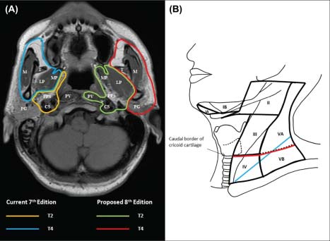
No change was recommended in the AJCC Cancer Staging Manual, 6th Edition5,6 except for addition of the term “masticator space” as a synonym for “infratemporal fossa” (one of the T4 criteria) because although the intended extent was described in the staging hand book, the latter was not a clearly defined space with universal acceptance. Both terms were retained as T4 criteria in the AJCC Cancer Staging Manual, 7th Edition (7th Edition);7,8 however, the term “masticator space” was described using the boundaries stated in classical anatomy textbooks instead of the demarcation used for “infratemporal fossa.” Additional changes included down-shifting of tumors with extension to nasal fossa/oropharynx without parapharyngeal extension (previously T2a) to T19 and clear definition of retropharyngeal lymph node(s) involvement (unilateral or bilateral) as N1.10

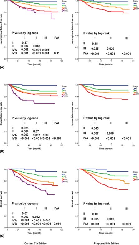
The management of NPC has undergone substantial evolution in the past two decades. More accurate imaging methods have allowed better delineation of tumor extent and early detection of occult metastases. The advances in radiotherapy technique has led to increasing conformity of tumor coverage and sparing of noninvolved structures. The use of combination chemotherapy has further improved tumor control and cure rates, especially for advanced locoregional disease. It is therefore important that the new staging system be based on data from patients managed with contemporary methods.

Extensive literature review showed that there are four major issues for consideration of improvement: (1) the controversy about the significance of “masticator space”,11-16 (2) uncertainty about the significance of prevertebral muscle invasion,17-19 (3) the possibility of replacing supraclavicular fossa (SCF)3 with anatomic nodal “levels,”20-25 and (4) simplification by elimination of unnecessary subgroups.25,26 These suggestions were validated by a large series of patients who were staged with magnetic resonance (MR) imaging and treated with intensity-modulated radiotherapy ± chemotherapy from two major centers (in Hong Kong and Fujian, China),27 before attaining consensus among international multidisciplinary experts. The strengths of the 7th Edition and the Chinese 2008 staging system23,24 are incorporated in developing the staging criteria in this AJCC Cancer Staging Manual, 8th Edition (8th Edition).

Anatomy

Primary Site(s)

The pharynx is divided into three regions: nasopharynx, oropharynx, and hypopharynx (Figure 9.1). The specific anatomic site of nasopharynx and regional lymphatics are described in this section.

The nasopharynx begins anteriorly at the posterior choana and extends along the plane of the airway to the level of the free border of the soft palate. It includes the superior wall, the posterior wall, and the lateral walls, which include the fossae of Rosenmuller and the mucosa covering the torus tubaris forming the Eustachian tube orifice. The floor is the superior surface of the soft palate. The posterior margins of the choanal orifices and of the nasal septum are included in the nasal fossa.

Nasopharyngeal tumors extending to the nasal cavity or oropharynx in the absence of parapharyngeal space involvement do not have a significantly worse outcome than tumors confined to the nasopharynx. Involvement of the parapharyngeal space is defined as posterolateral infiltration from the nasopharynx beyond the buccopharyngeal fascia into the triangular space lateral to the pharynx.

9.1 Anatomical sites and subsites of the nasopharynx, oropharynx, hypopharynx, and esophagus.

Regional Lymph Nodes

Nasopharyngeal carcinoma often presents with early lymphatic spread. The retropharyngeal nodes and the cervical nodes (both jugular and spinal accessory chains) are involved, often bilaterally. The lymphatic spread in NPC follows a predictable and orderly pattern from upper to lower neck; “skip” metastasis is rare.21,28

In clinical evaluation, the maximum dimension (in any direction) of the nodal mass, the laterality, and the lowest level of neck involvement should be assessed. Midline nodes are considered ipsilateral nodes. Nodal size larger than 6 cm in greatest dimension and /or extension below the caudal border of the cricoid cartilage are associated with the worst prognosis.

Metastatic Sites

Nasopharyngeal carcinoma is notorious for a high risk of distant metastasis. The most common sites include lung, bone, liver, and distant lymph nodes. Involvement of lymph nodes below the clavicle (including mediastinum, infraclavicular region, axilla, or groin) is considered as distant metastases.

Classification Rules

Clinical Classification

Clinical staging is employed for NPC. Assessment is based primarily on thorough history, physical examination, indirect or direct endoscopy, and imaging. Physical examination should include neurologic evaluation of all cranial nerves, palpation of neck nodes (greatest dimension, laterality, location, and lowest extent of nodal involvement), and exclusion of gross signs of distant metastases. Indirect or direct endoscopy should assess the extent of anterior involvement into the nasal cavities and inferior infiltration into the oropharynx and hypopharynx. Biopsy should be taken for histological confirmation. Routine testing for complete blood picture, renal, and liver functions (including alkaline phosphatase) are indicated.

Imaging

Cross-sectional imaging studies covering the nasopharyngeal and cervical regions are essential for clinical staging of NPC. Magnetic resonance (MR) imaging is the study of choice because of its multiplanar capability, superior soft tissue contrast, and sensitivity for detecting skull base and intracranial tumor spread. Computed tomography (CT) imaging with axial and coronal thin section technique with contrast is an alternative. Regional nodal status (greatest dimension in any direction, laterality, location, and lowest extent of nodal involvement) should be assessed; measurement of the maximal diameter of nodal disease should not be confined to the axial radiological plane only.

Metastatic workup is recommended for patients with node-positive or locally advanced (T3-4) disease, those with symptoms, signs, and /or biochemical tests suggestive of distant metastasis. Whole body 18F fluorodeoxyglucose (18F-FDG) positron emission tomography (PET) coupled with CT is increasingly used because of its sensitivity for detecting distant metastases and second primary malignancy, the possibility of its supplementing MR imaging in assessing nodal status,29 and its use of the maximal stand ard uptake values (SUVmax) as an additional independent prognostic predictor.30,31 Assessment by CT thorax and upper abdomen (or chest X-ray and abdominal ultrasound) and bone scan is an alternative.

Pathological Classification

Unlike other head and neck cancer, NPC is primarily treated by radiotherapy, with or without chemotherapy, with no resection of the primary cancer. This makes pathological classification largely irrelevant. Surgery to primary or neck nodes is used only for recurrence.

Prognostic Factors

Prognostic Factors Required for Stage Grouping

Although additional factors may contribute to refining prognostication, none have an adequate level of evidence and consistent cut-off value that attain consensus for incorporation as staging criteria.

Additional Factors Recommended for Clinical Care

Overall Health

In addition to the importance of the TNM factors, the overall health of these patients clearly influences outcome. An ongoing effort to better assess prognosis using both tumor and nontumor-related factors is underway. Chart abstraction will continue to be performed by cancer registrars to obtain important information regarding specific factors related to prognosis. These data will then be used to further hone the predictive power of the staging system in future revisions.

AJCC Level of Evidence: II

Comorbidity

Comorbidity can be classified by specific measures of additional medical illnesses. Accurate reporting of all illnesses in the patients' medical record is essential to assessment of these parameters. General performance measures are helpful in predicting survival. The AJCC strongly recommends that the clinician report performance status using the Eastern Cooperative Oncology Group (ECOG), Zubrod, or Karnofsky performance measures, along with stand ard staging information. An interrelationship between each of the major performance tools exists.

Zubrod/ECOG Performance Scale
0Fully active, able to carry out all predisease activities without restriction (Karnofsky 90-100)
1Restricted in physically strenuous activity but ambulatory and able to carry out work of a light or sedentary nature. For example, light housework, office work. (Karnofsky 70-80)
2Ambulatory and capable of all self-care but unable to carry out any work activities. Up and about more than 50% of waking hours. (Karnofsky 50-60)
3Capable of only limited self-care, confined to bed or chair 50% or more of waking hours (Karnofsky 30-40)
4Completely disabled. Cannot carry out self-care. Totally confined to bed. (Karnofsky 10-20)
5Death (Karnofsky 0)
AJCC Level of Evidence: II

Lifestyle Factors

Lifestyle factors such as tobacco and alcohol abuse negatively influence survival. Accurate recording of smoking in pack-years and alcohol in number of days drinking per week and number of drinks per day will provide important data for future analysis. Nutrition is important to prognosis and will be indirectly measured by weight loss of greater than 10% of body weight. Depression adversely impacts quality of life and survival. Notation of a previous or current diagnosis of depression should be recorded in the medical record.

The role of tobacco as a negative prognostic factor is well established. However, exactly how this could be codified in the staging system is less clear. At this time, smoking is known to have a deleterious effect on prognosis but is hard to accurately apply to the staging system. Smoking history should be collected as an important element of the demographics and may be included in Prognostic Groups in the future. For practicality, the minimum stand ard should classify smoking history as never, less than or equal to 10 pack-years, greater than 10 but less than or equal to 20 pack-years, or greater than 20 pack-years.

AJCC Level of Evidence: III

There are increasing reports that the following factors could aid in refining prognostication; however, they are AJCC Level of Evidence III and no consistent consensus yet exists on the demarcation criteria. These factors should be re-evaluated in the coming years as the evidence base grows.

  • Histological type (Nonkeratinizing versus keratinizing carcinoma)
  • Age
  • Gross tumor volume
  • Lactic dehydrogenase (LDH)
  • Circulating DNA of Epstein-Barr virus (EBV-DNA)

Risk Assessment

Risk Assesment Models

The AJCC recently established guidelines that will be used to evaluate published statistical prediction models for the purpose of granting endorsement for clinical use.32 Although this is a monumental step toward the goal of precision medicine, this work was published only very recently. Therefore, the existing models that have been published or may be in clinical use have not yet been evaluated for this cancer site by the Precision Medicine Core of the AJCC. In the future, the statistical prediction models for this cancer site will be evaluated, and those that meet all AJCC criteria will be endorsed.

Recommendations

As TNM staging remains the most important prognostic factor for NPC, it is recommended that clinical trials should at least be stratified by stage group.

TNM Definitions

Definition of Primary Tumor (T)

T CategoryT Criteria
TXPrimary tumor cannot be assessed
T0No tumor identified, but EBV-positive cervical node(s) involvement
TisTumor in situ
T1Tumor confined to nasopharynx, or extension to oropharynx and /or nasal cavity without parapharyngeal involvement
T2Tumor with extension to parapharyngeal space, and /or adjacent soft tissue involvement (medial pterygoid, lateral pterygoid, prevertebral muscles)
T3Tumor with infiltration of bony structures at skull base, cervical vertebra, pterygoid structures, and /or paranasal sinuses
T4Tumor with intracranial extension, involvement of cranial nerves, hypopharynx, orbit, parotid gland , and /or extensive soft tissue infiltration beyond the lateral surface of the lateral pterygoid muscle

Definition of Regional Lymph Node (N)

N CategoryN Criteria
NXRegional lymph nodes cannot be assessed
N0No regional lymph node metastasis
N1Unilateral metastasis in cervical lymph node(s) and /or unilateral or bilateral metastasis in retropharyngeal lymph node(s), 6 cm or smaller in greatest dimension, above the caudal border of cricoid cartilage
N2Bilateral metastasis in cervical lymph node(s), 6 cm or smaller in greatest dimension, above the caudal border of cricoid cartilage
N3Unilateral or bilateral metastasis in cervical lymph node(s), larger than 6 cm in greatest dimension, and /or extension below the caudal border of cricoid cartilage

Definition of Distant Metastasis (M)

M CategoryM Criteria
M0No distant metastatis
M1Distant metastasis

9.2 Differences in defining criteria between the 7th Edition and the 8th Edition for staging of NPC: (A) changing the extent of soft tissue involvement as T2 and T4 criteria. Abbreviation: CS = carotid space, LP = lateral pterygoid muscle, M = masseter muscle, MP = medial pterygoid muscle, PG = parotid gland , PPS = parapharyngeal space, PV = prevertebral muscle, T = temporalis muscle, (B) replacing supraclavicular fossa (blue) by lower neck, i.e., below caudal border of cricoid cartilage (red) as N3 criteria. From Pan et al.,27 with permission.

Stage Prognostic

!!Calculator!!

AJCC PROGNOSTIC STAGE GROUPS

When T is…and N is…and M is…Then the stage group is…
TisN0M00
T1N0M0I
T1, T0N1M0II
T2N0M0II
T2N1M0II
T1, T0N2M0III
T2N2M0III
T3N0M0III
T3N1M0III
T3N2M0III
T4N0M0IVA
T4N1M0IVA
T4N2M0IVA
Any TN3M0IVA
Any TAny NM1IVB

Registry Data

Registry Data Collection Variables

None

Histopathologic type

The World Health Organization (WHO) classification system33 is recommended for histopathologic classification, and the following histopathologic types are covered by the staging system (Table 9.1).

9.1 Classification of NPC

WHO classificationFormer terminology
Basaloid squamous cell carcinomaNo synonym exists (recently described)
Keratinizing squamous cell carcinomaWHO Type I (squamous cell carcinoma)
Nonkeratinizing carcinoma
DifferentiatedWHO Type II (transitional cell carcinoma)
UndifferentiatedWHO Type III (lymphoepithelial carcinoma)

Histologic grade

HISTOLOGIC GRADE (G)

A grading system is not used for NPCs.

Survival

9.3 Differences in prognostication of overall survival between the 7th Edition (Left) and the 8th Edition (Right) by (A) T-category, (B) N-category, and (C) stage group for NPC. From Pan et al., with permission.27

Bibliography

  1. Fleming I, Cooper J, Henson D, et al. American Joint Committee on Cancer. AJCC cancer staging manual. 5. Lippincott-Raven, Philadelphia; 1997.
  2. Sobin LH, Fleming ID. TNM Classification of Malignant Tumors, fifth edition (1997). Union Internationale Contre le Cancer and the American Joint Committee on Cancer. Cancer. Nov 1 1997;80(9):1803-1804.
  3. Ho J. Stage classification of nasopharyngeal carcinoma: a review. IARC scientific publications. 1977(20):99-113.
  4. Lee AW, Foo W, Law SC, et al. Staging of nasopharyngeal carcinoma: from Ho's to the new UICC system. Int J Cancer. Apr 20 1999;84(2):179-187.
  5. Greene FL. AJCC cancer staging manual. Vol 1: Springer Science & Business Media; 2002.
  6. Sobin LH WC. TNM classification of malignant tumours. International Union Against Cancer (UICC): 6th edition. New York: Wiley-Liss; 2002. 2002.
  7. Edge SB, Byrd DR, Compton CC, Fritz AG, Greene FL, Trotti A. AJCC cancer staging manual. Vol 649: Springer New York; 2010.
  8. Sobin L, Gospodarowicz M, Wittekind C. TNM classification of malignant tumors. Hoboken, NJ: Wiley-Blackwell; 2009.
  9. Lee AW, Au JS, Teo PM, et al. Staging of nasopharyngeal carcinoma: suggestions for improving the current UICC/AJCC Staging System. Clinical oncology. Jun 2004;16(4):269-276.
  10. Tang L, Li L, Mao Y, et al. Retropharyngeal lymph node metastasis in nasopharyngeal carcinoma detected by magnetic resonance imaging: prognostic value and staging categories. Cancer. Jul 15 2008;113(2):347-354.
  11. Tang LL, Li WF, Chen L, et al. Prognostic value and staging categories of anatomic masticator space involvement in nasopharyngeal carcinoma: a study of 924 cases with MR imaging. Radiology. Oct 2010;257(1):151-157.
  12. Chen L, Liu L-Z, Chen M, et al. Prognostic value of subclassification using MRI in the t4 classification nasopharyngeal carcinoma intensity-modulated radiotherapy treatment. International Journal of Radiation Oncology* Biology* Physics. 2012;84(1):196-202.
  13. Luo DH, Yang J, Qiu HZ, et al. A new T classification based on masticator space involvement in nasopharyngeal carcinoma: a study of 742 cases with magnetic resonance imaging. BMC cancer. 2014;14(1):653.
  14. Zhang GY, Huang Y, Cai XY, et al. Prognostic value of grading masticator space involvement in nasopharyngeal carcinoma according to MR imaging findings. Radiology. Oct 2014;273(1):136-143.
  15. Sze H, Chan LL, Ng W, et al. Should all nasopharyngeal carcinoma with masticator space involvement be staged as T4? Oral oncology. 2014;50(12):1188-1195.
  16. Xiao Y, Pan J, Chen Y, et al. The prognosis of nasopharyngeal carcinoma involving masticatory muscles: a retrospective analysis for revising T subclassifications. Medicine (Baltimore). Jan 2015;94(4):e420.
  17. Feng AC, Wu MC, Tsai SY, et al. Prevertebral muscle involvement in nasopharyngeal carcinoma. International journal of radiation oncology, biology, physics. Jul 15 2006;65(4):1026-1035.
  18. Lee CC, Chu ST, Chou P, Lee CC, Chen LF. The prognostic influence of prevertebral space involvement in nasopharyngeal carcinoma. Clin Otolaryngol. Oct 2008;33(5):442-449.
  19. Zhou GQ, Mao YP, Chen L, et al. Prognostic value of prevertebral space involvement in nasopharyngeal carcinoma based on intensity-modulated radiotherapy. International journal of radiation oncology, biology, physics. Mar 1 2012;82(3):1090-1097.
  20. Som PM, Curtin HD, Mancuso AA. Imaging-based nodal classification for evaluation of neck metastatic adenopathy. AJR. American journal of roentgenology. Mar 2000;174(3):837-844.
  21. Ng WT, Lee AW, Kan WK, et al. N-staging by magnetic resonance imaging for patients with nasopharyngeal carcinoma: pattern of nodal involvement by radiological levels. Radiotherapy and oncology : journal of the European Society for Therapeutic Radiology and Oncology. Jan 2007;82(1):70-75.
  22. Li W-F, Sun Y, Mao Y-P, et al. Proposed lymph node staging system using the International Consensus Guidelines for lymph node levels is predictive for nasopharyngeal carcinoma patients from endemic areas treated with intensity modulated radiation therapy. International Journal of Radiation Oncology* Biology* Physics. 2013;86(2):249-256.
  23. Pan J XY, Qiu S, et al. A Comparison Between the Chinese 2008 and the 7th Edition AJCC Staging Systems for Nasopharyngeal Carcinoma. American journal of clinical oncology. 2013;23:192-198.
  24. OuYang PY, Su Z, Ma XH, Mao YP, Liu MZ, Xie FY. Comparison of TNM staging systems for nasopharyngeal carcinoma, and proposal of a new staging system. Br J Cancer. Dec 10 2013;109(12):2987-2997.
  25. Yue D, Xu Y-F, Zhang F, et al. Is replacement of the supraclavicular fossa with the lower level classification based on magnetic resonance imaging beneficial in nasopharyngeal carcinoma? Radiotherapy and Oncology. 2014;113(1):108-114.
  26. Lee AW, Ng WT, Chan LK, et al. The strength/weakness of the AJCC/UICC staging system (7th edition) for nasopharyngeal cancer and suggestions for future improvement. Oral oncology. Oct 2012;48(10):1007-1013.
  27. Pan JJ, Ng WT, Zong JF, et al. Proposal for the 8th edition of the AJCC/UICC staging system for nasopharyngeal cancer in the era of intensity-modulated radiotherapy. Cancer. Nov 20 2015.
  28. Ho FC, Tham IW, Earnest A, Lee KM, Lu JJ. Patterns of regional lymph node metastasis of nasopharyngeal carcinoma: a meta-analysis of clinical evidence. BMC cancer. 2012;12(1):98.
  29. Ng SH, Chan SC, Yen TC, et al. Staging of untreated nasopharyngeal carcinoma with PET/CT: comparison with conventional imaging work-up. European journal of nuclear medicine and molecular imaging. Jan 2009;36(1):12-22.
  30. Lee S-w, Nam SY, Im KC, et al. Prediction of prognosis using stand ardized uptake value of 2-[18 F] fluoro-2-deoxy-d-glucose positron emission tomography for nasopharyngeal carcinomas. Radiotherapy and Oncology. 2008;87(2):211-216.
  31. Liu WS, Wu MF, Tseng HC, et al. The role of pretreatment FDG-PET in nasopharyngeal carcinoma treated with intensity-modulated radiotherapy. International journal of radiation oncology, biology, physics. Feb 1 2012;82(2):561-566.
  32. Kattan MW, Hess KR, Amin MB, et al. American Joint Committee on Cancer acceptance criteria for inclusion of risk models for individualized prognosis in the practice of precision medicine. CA: a cancer journal for clinicians. Jan 19 2016.
  33. Chan JKC PB, Kuo TT, Wenig BM, Lee AWM. Tumours of the nasopharynx. In: Eveson BL, JW RP, Sidransky D, editors. World Health Organization classification of tumour, pathology and genetics. Head and neck tumours. Lyon: IARC Press;. 2005:815-897.